Indications

Tumors in close proximity to radiosensitive organs are the main field of application of particle therapy. Pediatric tumors and local recurrences are also important patient groups in the treatment with charged particles.

This page provides information on all diseases that are currently treated at MedAustron.

Disease patterns and their treatment at MedAustron

  • Central Nervous System
  • Skull Base
  • Head & Neck
  • Gastrointestinal Tract
  • Pelvis
  • Sarcoma
  • Pediatric Malignancies
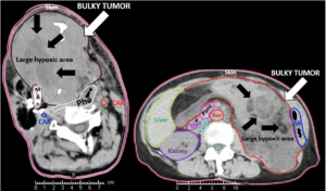
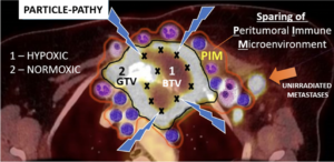
  • Unresectable Large / Bulky Tumors
1 — 8

Central Nervous System

Particle therapy, especially proton therapy, has been routinely performed for more than 30 years for various selective brain tumors as well as tumors of the skull base.

Particle therapy, i.e. proton and carbon ion radiotherapy, is primarily based on the better physical dose distribution, i.e. the radiation dose can be centered on the tumor, with a very fast decrease of the radiation dose towards the normal brain. Multiple comparisons with photon therapy have shown that the exposure of the normal brain to a low or moderate radiation dose can be significantly reduced with the use of protons and/or carbon ions. Even a relatively low radiation exposure of, however, larger parts of the normal brain can lead to long-term limitations of quality of life and neurocognitive functions. This is of course pronounced in childhood with a still developing brain and neurocognitive functions, which is why proton therapy is generally recommended for brain tumors. However, radiotherapy of the brain can also lead to considerable limitations in adult age, not only to the possible loss of specific functions, but also to general limitations of neurocognitive and intellectual resilience in professional but also social everyday life. In contrast, a prospective study at MedAustron showed preservation of all neurocognitive abilities for the entire group up to two years in >120 adult patients.

The advantage of particle therapy is not only evident in tumors that are exposed to relatively high radiation doses, but also in tumors that require relatively moderate doses (e.g. 50-54 Gy), but the volume to be irradiated is very irregularly shaped, especially towards the base of the skull.

Due to the sparing of normal brain tissue, particle therapy can also be used for dose escalation, i.e. to apply higher radiation doses to the tumor itself, in those tumors where local tumor control and thus generally the chances of survival are to be improved.

All patients are asked to participate in the registry study. This allows MedAustron to collect the data of the patients also in the context of aftercare, in order to be able to prove the effectiveness of particle therapy in the individual case and then also to be able to analyze the results (of course anonymized) for the whole group. Participation or non-participation in the registry study does not affect treatment in any way.

+
Tumors

The majority of pediatric and many adult CNS tumors requiring radiotherapy can be treated with particle therapy.

Proton therapy is part of the standard of care for the majority of pediatric brain tumors.

Increasingly, adult patients are also being assigned to particle therapy for specific indications.

+
Indications treated at MedAustron

Primary irradiations:

Low-grade gliomas (WHO I-III gliomas with evidence of an IDH1,2 mutation) but also other rare tumors within the CNS or in the direct vicinity such as pinaelis tumors, craniopharyngiomas, pituitary macroadenomas, ependymomas, schwannomas of the cranial nerves (e.g.: acoustic neuroma/vestibular schwannoma), meningiomas, hemangiopericytomas/solitary fibrous tumor, hemangioblastomas, and others.

According to the current state of science, high-grade gliomas (glioblastoma multiforme, grade IV, IDH neg. gliomas) benefit from particle therapy only in a few exceptions and highly individualized indications.

Re-irradiation:

Despite initially successful irradiation with photons (but possibly also after particle therapy), a certain percentage of patients may experience a recurrence of the tumor in the previous irradiation area or in the immediate vicinity.

The therapeutic procedure is discussed on a multi-disciplinary basis, i.e. whether the recurrence should best be removed surgically, whether systemic therapy, such as chemotherapy or immunotherapy, should be used, or whether irradiation should be applied again. If another course of irradiation is considered, either as a sole therapy or in combination, the question arises to what extent another full course of irradiation can be given, due to the prior exposure of normal tissue. In other words, do the brain structures, such as normal brain parenchyma or even important nerves, allow another radiation exposure in order to be able to treat this tumor with a necessary radiation dose?

Here, particle therapy can play a decisive role in enabling re-irradiation, which cannot be offered by conventional radiotherapy, i.e. many radiation oncologists justifiably deny re-irradiation with photons. However, a therapeutic decision is highly individual. In order to be able to make such a decision, a detailed review is required, especially of the irradiation plan that has already been applied.

MedAustron has developed various concepts involving either proton or carbon ion radiotherapy or a combination. Whether re-irradiation with particle therapy can be performed, and if so, with which concept, is decided by the medical team at MedAustron on an individual basis and discussed with the patient.

+
Example: Low Grade Glioma

22-year-old female patient with a low grade glioma (WHO II, IDH mut) in the left opercular region of the left hemisphere.

Underwent biopsy and partial resection. Complete surgery not feasible. Sent for Proton Therapy.

Treated according to institutional protocol protocol to total dose of 54 Gy (RBE).

Example CNS Indication: Low Grade Glioma

Low Grade Glioma (click to enlarge image)

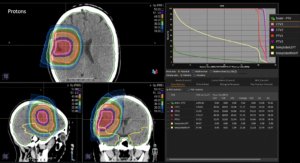
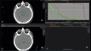
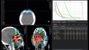
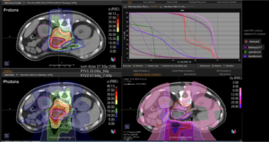
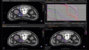
Graph and video demonstrating the full irradiation plan using proton therapy:

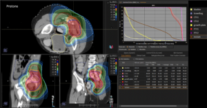
Left side: Proton plan in 3 views: axial, sagittal (side) and coronal (frontal view).
Right side: dose/volume histograms (DVH‘s), meaning the graphic distribution of dose delivered to the volume of specific organs – in this case brain and temporal lobes. In addition numerical display of various dose levels.

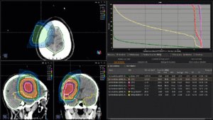
Low grade glioma treatment plan

Low grade glioma treatment plan (click to enlarge image)

Animation: low grade glioma treatment plan

Animation: low grade glioma treatment plan (click to start and enlarge animation)

Note: Of clinical importance was achieving an optimal tumor coverage, despite not exceeding doses to the critical organs located in direct proximity: the non-involved healthy CNS parenchyma especially the temporal lobe on both sides. Note the complete absence of dose to the contralateral brain.

The excellent dose distribution is expected to result in a low risk of brain toxicity and low risk of decline of cognitive function.

+
Side Effects

In general, there are no side effects specific to particle therapy, but side effects may occur, as with conventional photon therapy, too. Since less dose reaches the surrounding normal tissue due to the physical properties described, these side effects can be significantly less pronounced.

Because CNS tissue can be very sensitive to ionizing radiation (radiotherapy) and thus be damaged in the long term. One of the main causes is damage to the smallest blood vessels/capillaries, which in turn leads in the long term to a reduction in blood circulation and corresponding damage to the tissue. These effects can also be observed in areas that received only a low dose.

Clinically, this can lead to neurocognitive changes and mental slowing. Further side effects of radiotherapy, depending on the localization, can be neurological changes up to deficits (hearing, visual acuity, sense of taste/smell,…) as well as hormonal changes / deficits if the pituitary gland is involved. All of these effects can occur years after radiotherapy and affect quality of life. The risk of these late effects can be significantly reduced with proton therapy.

+
FAQ

a. Can particle therapy be combined with systemic therapy?
i. Depending on the tumor, particle therapy can be combined with systemic therapy, as it is with conventional radiotherapy as well. In such cases, the indication is made on an interdisciplinary basis (medical oncologist and radiation oncologist).

b. Is particle therapy possible after surgery as well?
i. Particle therapy can also be performed after surgery, especially if the surgeon could not remove everything, it is also indicated.

c. Is there a limitation on the tumor size in particle therapy?
i. In principle, tumors of any size can be treated with particle therapy.

d. Can particle therapy replace surgery?
i. If a tumor is accessible to surgery, this is the treatment of choice in the majority of cases. In some situations, surgery is associated with high risk, so that irradiation can be performed as an option with curative intent.

e. Can particle therapy cause side effects?
i. As with conventional irradiation, local reactions can also occur in the surrounding normal tissue – but as a rule these are much less pronounced, since due to the physical properties this tissue is exposed to a lower dose.

f. Are the chances of cure higher after particle therapy than after conventional irradiation?
i. The biological effect of proton therapy is very similar to that of photon therapy; thus, the chances of cure at the same dose are identical after proton therapy as after conventional irradiation. Due to the lower normal tissue exposure, in special situations the total dose can be increased during proton therapy and consequently an improvement in local control can be achieved.

g. Are there age limits for particle therapy?
i. In principle, patients of any age who are indicated for radiotherapy can be treated with particle therapy.

+
Literature

1. Superior Intellectual Outcomes After Proton Radiotherapy Compared With Photon Radiotherapy for Pediatric Medulloblastoma. Kahalley LS, Peterson R, Ris MD, et al. J Clin Oncol. 2020 Feb 10;38(5):454-461.

2. Proton therapy for selected low grade glioma patients in the Netherlands. van der Weide HL, Kramer MCA, Scandurra D, et al. Radiother Oncol. 2021 Jan;154:283-290.

3. Proton therapy for treatment of intracranial benign tumors in adults: A systematic review. Lesueur P, Calugaru V, Nauraye C, et al. Cancer Treat Rev. 2019 Jan;72:56-64.

4. Proton Therapy for Intracranial Meningioma for the Treatment of Primary/Recurrent Disease Including Re-Irradiation. Weber DC, Bizzocchi N, Bolsi A, et al. Front Oncol. 2020 Dec 14;10:558845.

5. A Multi-institutional Comparative Analysis of Proton and Photon Therapy-Induced Hematologic Toxicity in Patients With Medulloblastoma. Liu KX, Ioakeim-Ioannidou M, Susko MS, et al. Int J Radiat Oncol Biol Phys. 2021 Mar 1;109(3):726-735.

6. A Systematic Review on Re-irradiation with Charged Particle Beam Therapy in the Management of Locally Recurrent Skull Base and Head and Neck Tumors. Gamez ME, Patel SH, McGee LA, et al. Int J Part Ther. 2021 Jun 25;8(1):131-154.

7. Re-irradiation with protons or heavy ions with focus on head and neck, skull base and brain malignancies. Seidensaal K, Harrabi SB, Uhl M, et al. Br J Radiol. 2020 Mar;93(1107):20190516

2 — 8

Skull Base

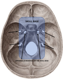
From an anatomical point of view, the skull base is represented by a group of bones that have fused together to form the base of the skull on which the brain is laid (see below Fig. 1).

Practically, the base of the skull separates the brain and other intracranial organs and tissues from the extracranial ones with which they communicate through numerous cavities and openings in the bones which contain blood vessels or nerves for example. Through these cavities also the tumor can be drawn inside the skull reaching and infiltrating the skull base and all those important healthy tissues and organs.

Skull Base

Fig.1: Skull base region: the area highlighted in blue corresponds to the region where the group of the skull bones meet together to form the base of the skull on which the brain is laid. At this level, several bone apertures or foramina are observable bilaterally through which different cranial nerves and the vessels entering or exiting the skull (click to enlarge image).

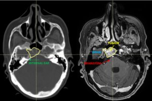
It is that very close and intimate relationship of the skull base region with very important organs and tissues that pass there that makes the tumors of this region very difficult and complicated to treat (Fig.2). That´s why one of the typical indications for particle therapy are the skull base tumors. Particle therapy, especially proton therapy, is routinely used for more than 30 years in the treatment of different skull base tumors. The reason for that is a superior and more favorable radiation dose distribution of the particles compared to the photons (standard, conventional radiation) whereby the higher radiation dose can more safely be delivered to the tumor while the surrounding healthy tissue structures and organs can better be spared. That saying, the probability for tumor control potentially increases while for the side effects decreases with particle therapy. This is possible thanks to the special physical proprieties of the particles, both protons and carbon ions, which are endowed by a mass and charge, both determining the sharper dose fall off outside the tumor, at the level of the organs at risk. This is particularly true for the organs of the critical importance for vision, hearing or cognitive, motoric and sensitive functions like optical nerves, internal ears, basal ganglia or brainstem, all they usually infiltrated or compressed by the skull base tumors. The conventional radiotherapy using photons, that are the electromagnetic fields without the mass and charge, cannot reach the required, high enough tumoricidal radiation dose necessary to eradicate and control certain skull base tumor without exciding dose-constraints of those nearby critical healthy organs.

Skull base tumor diagnostic representation

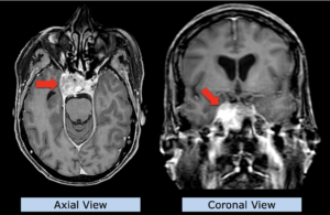
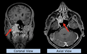
Fig. 2: Diagnostic representation of a skull base tumor (yellow contour): in planning magnetic resonance (left) and in computer tomography (right) (click to enlarge image).

+
Indications treated at MedAustron

For the great majority of skull base tumors, radiotherapy plays an essential role either as a primary treatment for unresectable tumors or as an irreplaceable element of multidisciplinary approach for those tumors that are partially resectable.

For chordomas and chondrosarcomas the standard of care is maximum safe surgical resection followed by particle therapy. For this tumor the treatment strategy should be discussed upfront with the surgeon and the radiation oncologist so that the combined treatment ( surgery + postoperative particles) is planned simultaneously.

For pituitary adenomas, schwannomas (neuromas) of various cranial nerves including acoustic neuromas, optic pathway gliomas, paragangliomas and esthesioneuroblastomas particle therapy can be used as exclusive treatment. Each case must be evaluated singularly, together with the neurosurgeon. Particle therapy can be used as first treatment option when radical surgery is either not possible or too mutilating. In addition to all these primary tumors originating from skull base region, many others either primary outside the skull base region or secondary (metastatic) tumors from very distant body regions that involve the skull base are also considered for particle therapy at MedAustron. These include the primary head and neck tumors extending into the skull base, like for ex. paranasal sinus tumors or adenoid cystic carcinomas.

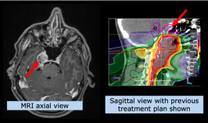
Re-irradiation after local failure of conventional radiotherapy is also added to our list of indications on an individual basis. Despite initially successful photon irradiation (but possibly also after particle therapy), a certain percentage of patients may experience a local recurrence of the tumor in the previous area of irradiation, or in the immediate vicinity (also called “relapse”).

The therapeutic approach is discussed in a multidisciplinary manner, i.e. whether the relapse should best be removed surgically which is rarely possible, whether systemic therapy, such as chemotherapy or immunotherapy, should be used especially for tumor-reduction, or even repeated radiation. Should a further irradiation either as the sole therapy or in combination with other therapeutic options be considered, the question arises if a full (curative) dose of irradiation can be given again, due to the previous exposure of normal tissues. In other words, the previously received radiation dose by the normal brain structures, such as brain parenchyma or cranial nerves, is critical in determining a possibility of subsequent re-exposure to radiation. Here, the previously mentioned characteristics of particle therapy can make a decisive contribution to enabling re-irradiation that conventional radiation therapy can no longer offer, and thus would be otherwise denied with photons. A therapy decision, however, is highly individual. In order to be able to make such a decision, a detailed review is required, especially of the radiation plan that has already been applied.

MedAustron has developed various concepts that include either proton therapy or carbon ion therapy or a combination of those two. Whether a re-irradiation with particle therapy can be carried out and if so, with which concept, is decided individually by the medical team at MedAustron and discussed with the patient.

+
Example: Skull Base Sarcoma

75-year-old woman with skull base sarcoma.

She underwent biopsy only, since complete surgical resection was not considered feasible.

She underwent a full course of Carbon Ion Radiotherapy and was treated to a total dose of 76.8 Gy (RBE) over 4 weeks in 16 treatment fractions (hypofractionated).
Example: skull base sarcoma

Skull base sarcoma (click to enlarge image)

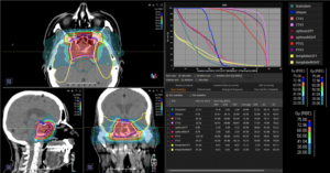
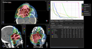
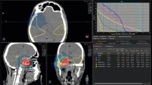
Graph and video demonstrating the full irradiation plan using CARBON ION therapy:

Left side: Carbon ion plan in 3 views: axial, sagittal (side) and coronal (frontal) view.

Right side: Dose/volume histograms (DVH‘s), meaning the graphic distribution of dose delivered to the volume of specific organs – in this case brainstem, optic chiasm, optic nerves and temporal lobes. In addition numerical display of various dose levels.

Treatment plan skull base sarcoma

Treatment plan skull base sarcoma (click to enlarge image)

Animation: treatment plan skull base sarcoma

Animation: treatment plan skull base sarcoma (click to start and enlarge animation)

Note: Of clinical importance is the high degree of conformality or accuracy of high doses around the target volume – yet the sharp dose fall-off towards the normal brain (in this case the temporal lobes). Despite the immediate proximity of tumor, the dose could be limited to safe dose levels towards the optic chiasm, both optic nerves and the brainstem surface.

+
Side Effects

Generally considered, there are no particle therapy-specific side effects. As it happens also by using a conventional photon radiotherapy, side effects can occur even by particle therapy. Since, due to the previously described physical properties of particles, less radiation dose reaches the surrounding normal tissues, these side effects are however significantly less pronounced. Since the intracranial tissues surrounding the skull base area are not resistant to the ionizing radiation, they can be damaged during particle therapy even on the long term basis. One of the main reasons for this is the radiation-induced damage to the smallest blood vessels/capillaries, which in turn leads to a decrease in blood flow over the long term resulting in damage of the healthy tissues. These changes can also be seen in the areas that received a low radiation dose. Clinically, this can lead to neurocognitive changes and mental slowdown. Finally, other side effects of radiation include but are not limited to the neurological deficits or even hearing-, vision-, taste/smell-loss and, if the pituitary gland is involved, hormonal changes in terms of pituitary insufficiency. All of these changes can occur even years after radiation therapy and affect quality of life. The risk of these long-term effects can be significantly reduced with proton irradiation.

+
FAQ

a. Can particle therapy be combined with systemic therapy?
i. Depending on the tumor type, particle therapy, as with conventional radiation therapy, can be combined with systemic therapy. In such a case, the indication is made on an interdisciplinary basis (medical oncologist and radiation oncologist).

b. Is particle therapy also possible following an operation?
i. Particle therapy can also be carried out after an operation, especially if the surgeon was unable to remove everything, this is indicated.

c. Is there a size limit for the tumor to be treated with particle therapy?
i. In principle, tumors of any size can be treated with particle therapy.

d. Can particle therapy replace surgery?
i. If a tumor is amenable to surgery, this is usually the treatment of choice. In some situations, an operation is associated with a high risk, so that, as an alternative, particle therapy can be carried out with curative intent.

e. Can there be side effects of particle therapy?
i. With conventional irradiation, reactions can also occur locally in the surrounding normal tissue – but these are usually much less pronounced compared to conventional radiotherapy, as these tissues are exposed to a lower radiation dose due to the physical properties of particles.

f. Are the chances of recovery higher after particle therapy than after conventional radiation?
i. The biological effect of proton therapy is very similar to that of photon therapy, thus the chances of recovery are the same after proton therapy as after conventional radiation with the same dose. Due to the improved normal tissue sparing, the total dose for proton therapy can be increased in certain situations, leading consequently to an improvement in local tumor control.

g. Are there age limits for particle therapy?
i. In principle, patients of any age for whom radiation is indicated can be treated with particle therapy.

+
Literature

1. Systematic Review on Re-irradiation with Charged Particle Beam Therapy in the Management of Locally Recurrent Skull Base and Head and Neck Tumors. Gamez ME, Patel SH, McGee LA, et al. Int J Part Ther. 2021 Jun 25;8(1):131-154.

2. Re-irradiation with protons or heavy ions with focus on head and neck, skull base and brain malignancies. Seidensaal K, Harrabi SB, Uhl M, et al. Br J Radiol. 2020 Mar;93(1107):20190516.

3. A Review of Particle Therapy for Skull Base Tumors: Modern Considerations and Future Directions. Eugen B. Hug, MD; Maciej Pelak, MD, PhD; Steven J. Frank, MD; Piero Fossati, PhD Int J Part Ther (2021) 8 (1): 168–178.

3 — 8

Head & Neck

Tumors of the head and neck region are particularly challenging for radiation oncology. This anatomic area is relatively small but densely filled with important, sensitive healthy organs that can be only millimeters away from the tumor.

The outcome of local treatment, both in terms of achieving long-term tumor control and keeping the toxicity acceptable, has a key influence on quality of patients’ life. For many tumor types and locations within head and neck region the particle therapy can successfully achieve both optimal tumor irradiation and protection of organs at risk, often challenging for conventional therapy. As of 2021, tumors of the head and neck region are the most frequent of all indications treated at MedAustron.

The clinical advantage of particle therapy is based on the physical properties of proton and ion beams, including low entry dose, ability to precisely align maximal dose of each beam at the tumor depth and either no (for protons) or minimal (for carbon ions) exit dose. Additionally, carbon ions, due to their biologic properties as heavy ions, produce more significant damage to the tumor cells when compared with photons and protons. This property is used to successfully treat tumors which are known to feature increased resistance to conventional radiation. For tumors which require a total radiation dose of approximately 70 Gray or less, proton therapy delivers tumor control which is comparable to that of conventional therapy, but usually with significantly fewer side effects. For tumors that require higher doses, up to 78 Gray, particle therapy is even more strongly indicated because the conventional radiation can often not deliver such doses to the tumors in the head and neck region without risking severe side effects.

Most head and neck tumors feature a low to moderate risk of spreading to other organs after the primary treatment compared with other tumor types (i.e. lung or breast cancer), therefore an effective and safe local therapy is of paramount importance for improved patient survival and maintaining good quality of life.

All head & neck patients at MedAustron are encouraged to take part in the registry study, in which tumor control and side effects are prospectively recorded. This enables us to assess and publish, after anonymization, the outcomes of the therapy we offer. The participation is not mandatory, carries no additional risks or obligations and the treatment received does not differ for patients participating or not in the registry study.

+
Tumors

The majority of tumors of the head & neck can be treated with particle therapy, however patients have the greatest benefit in comparison to conventional radiation for tumors that are located close to orbit, optic nerves, optic chiasm, brain stem or inner ears – each of these organs has a threshold of radiation dose which should not be exceeded in order to avoid unacceptable risk of severe chronic side effects associated with these organs.

Tumors of rare histological types known to feature increased resistance to radiation usually profit from the particle therapy regardless of their specific location within the head & neck region, in particular for inoperable or incompletely resected disease. This specifically applies for patients in whom re-irradiation (= a second course of radiotherapy in the same anatomic area) is indicated.

Proton therapy can be used for the same indications as conventional radiation – both in primary and postoperative treatment including cases of prophylactic irradiation with no macroscopic tumor present, also with simultaneous chemotherapy if indicated. Irradiation with carbon ions is currently only indicated when macroscopic disease is present, either in inoperable or incompletely resected cases. It can be coupled with concomitant systemic treatment.

An innovative approach at MedAustron combines proton with carbon ion therapy.

+
Indications treated at MedAustron

Primary irradiation:

• Malignant tumors of nasal cavity and paranasal sinuses
• Tumors of salivary glands (in particular adenoid-cystic carcinoma)
• Tumors of oropharynx and nasopharynx with skull base involvement
• Certain tumors of the orbit (i.e. rhabdomyosarcoma, primary tumors of lacrimal gland)
• Tumors of external auditory canal and middle ear

For selected clinically benign lesions (glomus tumors, peripheral neurinomas) proton therapy is also indicated and offered at MedAustron.

In primary treatment of tumors of the tongue, oral cavity, larynx, hypopharynx, thyroid or skin, patients are treated in individual cases. Patients should be referred by the interdisciplinary tumor board of the institution where the tumor was diagnosed and/or operated.

Re-Irradiation:

Just like any other tumor in the human body, tumors of the head and neck region can recur or progress after initial treatment with radiation. Additionally, radiation itself can induce tumor growth in healthy tissue in rare instances. Secondary, radiation-induced tumors appear later than recurrences of the same tumor and usually are of different histological type. Because of a high dose already delivered to the same area, a great majority of these newly developed tumors are particularly challenging to treat.

Re-irradiations are a frequent indication at MedAustron, second in numbers only to primary head & neck treatment. The feasibility of this treatment has to be carefully evaluated, taking into account the doses previously delivered to the organs at risk, tolerance of the previous treatment and any of its persistent side effects, general patient condition, alternative treatments available a.o. At MedAustron we have developed strategies to treat recurrent tumors according to their histology, location and other oncological considerations. Compared to tumors with no prior radiation treatment, re-irradiation is often associated with a lower chance of tumor control and higher toxicity but is still an invaluable option for patients who are not candidates for surgery. To overcome potential resistance of recurrent tumors to radiation it is often delivered with a higher dose per fraction in accelerated schemes which take between 1 and 4.5 weeks to complete (compared to 4-7.5 weeks for primary treatment).

Any recurrent tumor in the head & neck region can be considered for re-irradiation at MedAustron as in majority of cases these benefit from lower dose to the organs at risk and high efficacy against the tumor. The decision is highly individualized. In order to assess the feasibility for this treatment we require to have:
• Current clinical status, in particularly regarding the local symptoms
• Previous irradiation plans, preferably digital if available
• Current imaging studies (CT, MRI, PET/CT)

+
Example: Pleomorphic Adenoma

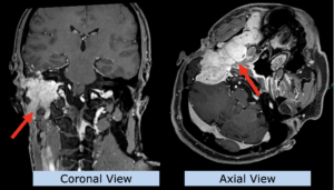
A 51-yea- old patient with a history of multiple local recurrences after surgical resections of a pleomorphic adenoma, a locally aggressive tumor of the right parotid gland.

The last recurrence was extensive and not feasible for surgery.

Irradiated with curative intent with CARBON IONS to a total dose of 68.8 Gry(RBE) in 16 fractions over 4 weeks (hypofractionated).
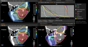
Example head and neck: adenoma of parotid gland

Pleomorphic adenoma of parotid gland (click to enlarge image)

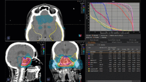
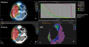
Planning Comparison between modern Photon Therapy (VMAT) with Proton Therapy at MedAustron:

Left upper panel photon plan, left lower panel proton plan.
Right upper panel dose/volume histogram, meaning the graphic distribution of dose delivered to the volume of specific organs
Right lower panel the „differential dose“ – meaning the additional dose delivered to normal tissues and structures by photons in comparison to protons , that does not contribute to tumor dose.

Plan comparison adenoma of parotid gland

Plan comparison pleomorphic adenoma of parotid gland (click to enlarge image)

Animation: plan comparison adenoma of parotid gland

Animation: plan comparison pleomorphic adenoma of parotid gland (click to start and enlarge animation)

Note: Of clinical importance were significantly reduced dose delivered to the whole volume of oral cavity (= improved tolerance, reduction of pain and swallowing difficulties during treatment by leaving the opposite side of the mouth free of mucosal inflammation) and mobile tongue (= improved tolerance during treatment, reduced pain and reduced risk of chronic impairment of taste).
Additionally, the particle therapy reduces unneccessary radiation dose to normal brain, not involved by the tumor in this patient.

In this particular case an additional advantage of carbon ions in achieving permanent tumor control and cure can be expected as this rare tumor type is known to be resistant to conventional photon radiation.

+
Example: Adenocarcinoma of Maxillary Sinus

A 65-year-old patient with a newly diagnosed adenocarcinoma of the right maxillary sinus, extending into the palate and along the cranial nerves.

Disease extent considered inoperable (even an extensive surgery would unlikely result in complete removal of the tumor).

Irradiated with curative intent with CARBON IONS to a total dose of 68.8 Gy(RBE) in 16 fractions over 4 weeks.

Example head and neck: adenocarcinoma

Adenocarcinoma of Maxillary Sinus (click to enlarge image)

Graph and video demonstrating the full re-irradiation plan using CARBON ION therapy:

Left side: Carbon ion plan in 3 views: axial, sagittal (side) and coronal (frontal) view.

Right side: Dose/volume histograms (DVH‘s), meaning the graphic distribution of dose delivered to the volume of specific organs – in this case brainstem, cochlea, contralateral parotid, temporal lobes. In addition numerical display of various dose levels.

Treatment plan adenocarcinoma

Treatment plan adenocarcinoma of maxillary sinus (click to enlarge image)

Animation: treatment plan adenocarcinoma

Animation: treatment plan adenocarcinoma of maxillary sinus (click to start and enlarge animation)

Note: Of clinical importance was achieving an optimal tumor coverage, despite not exceeding doses to the critical organs located in direct proximity: the inner ear, the temporal lobe on the involved side and brain stem. Thanks to this, a low risk of complications associated with these organs was achieved.

In addition maximum sparing of dose to the entire oral cavity and contralateral parotid gland. This improved treatment tolerance by reducing pain and swallowing difficulties. The low dose to contralateral Parotid gland ascertained sufficent saliva production to avoid a permanently dry mouth.

An additional advantage of carbon ions in achieving tumor control can be expected as this tumor type is known to be resistant to conventional photon radiation.

+
Example: Nasopharynx Carcinoma

A 42-year-old patient was treated previously with concomitant chemo- and radiotherapy up to 70 Gy for Cancer of the Nasopharynx. One year later a recurrence was identified at the upper border of the previously treated area.

Due to the location in the skull base and involvement of the right trigeminal nerve it was considered unresectable.

He received a second, full course of proton therapy with a curative intent (=Re-irradiation) to a total dose of 66 Gy (RBE) in 22 fractions over 4.5 weeks.

Example Head and Neck: Nasopharynx Carcinoma

Nasopharynx carcinoma (click to enlarge image)

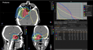
Graph and video demonstrating the full re-irradiation plan using proton therapy:

Left side: Proton plan in 3 views: axial, sagittal (side) and coronal (frontal view).
Right side: dose/volume histograms (DVH‘s), meaning the graphic distribution of dose delivered to the volume of specific organs – in this case optic chiasm, optic nerve, temporal lobes. In addition numerical display of various dose levels.

Treatment plan nasopharynx carcinoma

Treatment plan nasopharynx carcinoma (click to enlarge image)

Animation: treatment plan nasopharynx carcinoma

Animation: treatment plan nasopharynx carcinoma (click to start and enlarge animation)

Note: The doses previously received by healthy organs located next to the tumor recurrence made it challenging to deliver the required dose to the tumor. Of critical importance was not exceeding a maximum dose of 41 Gy (RBE) to the brainstem surface in the new treatment plan, which would have lead to increased risk of potentially life-threatening side effects from this organ. The optic chiasm also had to be protected in order to decrease the risk of bilateral vision loss.

+
Side Effects

The side effects of particle therapy are of similar type to those caused by conventional radiation, however their severity and frequency is usually lower. The majority of patients undergo from start to finish particle therapy on an outpatient basis, commuting from home or staying at a hotel or apartments nearby. If, however, the symptoms become severe and need supportive therapy that can only be given in a hospital, we continue treatment with the patient admitted to our collaborating division – Head & Neck Department of Landesklinikum Wiener Neustadt. This is a relatively rare situation among our patients and mostly concerns those patients in whom coupling of radiation with simultaneous chemotherapy is indicated.

Different organs respond to radiation in different ways. Two types of side effects have to be expected, depending on the anatomic location and dose prescription:

Early side effects – occurring during treatment or shortly thereafter, are quite frequent among patients but well controllable with medication and with a tendency to heal within weeks or completely resolve within a few months after treatment completion:
• Local skin reaction (from reddish like a sunburn up to superficial wounds)
• Local mucosal reaction (sore spots in the mouth, pain, obstructed nose, superficial bleeding, etc.)
• Dry mouth or excess of thick saliva
• Red, dry or watery eye
• Temporary loss or changing taste and smell
• Pain during swallowing, loss of appetite
• Hearing deterioration (accumulation of fluid in middle ear), ear pain
• Subcutaneous edema (swelling of skin)

Late side effects – typically occurring 3 months or later after treatment completion. They are rare (up to 5% based on historic data), however their treatment can become challenging and can remain chronic:
• Impaired vision and hearing
• Chronic dry mouth, loss of taste
• Hardening of skin, chronic oedema
• Trismus (restriction of mouth opening)
• Chronic sinonasal inflammation (in particular in patients after multiple surgeries in this area)
• Cataract
• Hormonal deficits (pituitary & thyroid)
• Others depending on organs at risk adjacent to irradiated site

+
Literature

1. Jingu K., Tsuji I., Mizoe J. et al. Carbon Ion Radiation Therapy Improves the Prognosis of Unresectable Adult Bone and Soft-Tissue Sarcoma of the Head and Neck. Int J Radiat Oncol Biol Phys. 2011; 82: 2125-31. doi: 10.1016/j.ijrobp.2010.08.043.

2. Mizoe J.-E., Hagesawa A., Jingu K. et al. Results of Carbon Ion Radiotherapy for Head and Neck Cancer. Radiother Oncol. 2012; 103: 32-37. doi: 10.1016/j.radonc.2011.12.013.

3. Sulaiman N.S., Demizu Y., Koto M. et al. Multicenter Study of Carbon-Ion Radiation Therapy for Adenoid Cystic Carcinoma of the Head and Neck: Subanalysis of the Japan Carbon-Ion Radiation. Oncology Study Group (J-CROS) Study. Int J Radiat Oncol Biol Phys. 2018; 100: 639-646. doi: 10.1016/j.ijrobp.2017.11.010.

4. Yu N.Y., Gamez M.E., Hartsell W.F. et al. A Multi-Institutional Experience of Proton Beam Therapy for Sinonasal Tumors. Adv Radiat Oncol. 2019;4(4):689-698. doi:10.1016/j.adro.2019.07.008

5. Pelak M.J., Walser M., Bachtiary B. et al. Clinical outcomes of head and neck adenoid cystic carcinoma patients treated with pencil beam-scanning proton therapy. Oral Oncol. 2020;107:104752. doi: 10.1016/j.oraloncology.2020.104752.

6. Chowdhury I. Nead K.T., Lustig R.A. et al. First Report of Paragangliomas Treated With Proton Therapy. Int J Radiat Oncol Biol Phys. 2018;96(S2):E126 doi:10.1016/j.ijrobp.2016.06.907

7. McDonald MW, Liu Y, Moore MG, Johnstone PA. Acute toxicity in comprehensive head and neck radiation for nasopharynx and paranasal sinus cancers: cohort comparison of 3D conformal proton therapy and intensity modulated radiation therapy. Radiat Oncol. 2016;11:32. Published 2016 Feb 27. doi:10.1186/s13014-016-0600-3

4 — 8

Gastrointestinal Tract

Tumors of the gastrointestinal tract must be treated with a multidisciplinary approach. In all cases a multidisciplinary evaluation is necessary and the patient must be discussed at least by a surgeon, a medical oncologist, a radiation oncologist and a diagnostic radiologist.

The optimal treatment must be decided for each individual patient. Particle therapy is a kind of radiotherapy and as such a local treatment. It can be used before surgery, after surgery or instead of surgery. For tumors of the liver, several other non-surgical local therapies are possible and particle therapy has to be integrated in this complex scenario.
In the thorax and upper abdomen there are several organs at risk such as heart, lungs, small intestine, stomach, liver, and kidneys that may be damaged by radiation and cause unwanted side effects.

Particle therapy with both protons and carbon ions has been used since more than 20 years in the treatment of gastrointestinal tumors. Both particles can achieve an optimal sparing of the noninvolved healthy tissues and therefore potentially reduce unwanted side effects and toxicity in comparison with photons based radiotherapy.

Because of the advantage in dose distribution, particle therapy can also in some case allow a safe escalation of the dose and improve the chance of local control. This higher probability of local control can be achieved prescribing a higher dose of proton therapy or taking advantage of the high relative biological efficacy of carbon ions radiotherapy.

+
Tumors

Tumors of the gastrointestinal tract can be divided in tumor originating from hollow and solid organs.

The hollow organs are: esophagus, stomach, small intestine and large intestine. The liver and pancreas are the solid organs of the digestive system.

Hollow organs have a wall that is typically sensitive to radiation damage. Exceeding the tolerance dose of these organs can result in severe toxicity such as ulceration, bleeding, and perforation. Because of these reasons no kind of radiotherapy (including particle therapy) is used in the treatment of small and large intestinal tumors. Stomach tumors are treated with radiotherapy, but the stomach itself is the most critical organ at risk and therefore there is little theoretical benefit in particle therapy over photons. Particle therapy can play a role in the treatment of primary tumors of the liver, of the pancreas and of the biliary tract. Particle therapy can play a role in the treatment of esophageal tumor. The esophagus is a hollow organ and therefore, even with particle therapy, dose escalation is not possible as the esophagus itself limits the dose that can be delivered. However, particle therapy can be used to treat esophageal tumors because by improving sparing of heart and lungs it can significantly reduce the risk of toxicity. Particle therapy is not used for the first line treatment of rectal cancer but can play a role in the treatment of local recurrences.

When the primary tumor has already spread through the blood (for example a pancreatic tumor that has caused metastasis in the liver) no local treatment can be curative.

Metastatic patients can still achieve a durable tumor response and a prolonged survival but this is possible thanks to systemic treatment such as chemotherapy or modern targeted drugs. The local treatment of metastatic disease can still have an important symptomatic role and improve the quality of life. In the vast majority of cases, the symptomatic irradiation of metastatic disease can be performed with photons based radiotherapy and particle therapy is not employed.

+
Indications treated at MedAustron

1. Pancreatic cancer

A minority of pancreatic cancers arise from the hormone producing part of the gland (the so-called endocrine pancreas). These tumors (insulinoma, gastrinoma, glucagonoma, pancreatic NET or neuroendocrine tumors, etc.) have a relatively good prognosis and can be treated with surgery, and other medical therapies. Particle therapy plays no role in the treatment of endocrine pancreatic tumors.

The majority of pancreatic tumors however originates from the portion of the gland that produces the digestive juices. These so-called exocrine pancreatic tumors are more difficult to treat and have a worse prognosis.

Unfortunately almost half of the exocrine pancreatic tumor are already metastatic at the diagnosis. The most common sites of metastasis are the liver and the peritoneum (the so-called peritoneal carcinosis). These patients must be treated with systemic therapies and particle therapy plays no role in metastatic pancreatic cancer.

A minority of patients are diagnosed in an early stage and are candidate to upfront radical surgery. These patients should proceed to surgery without delay.

There is about one third of the patients that, at the time of diagnosis, are not metastatic but have a disease, which cannot be resected surgically. This is often the case because the pancreas is located close to important blood vessels that cannot be resected and pancreatic tumors tend to invade these vessels. According to the importance of the vessel and the degree of invasion these patients are classified as “borderline resectable” or as “locally advanced” and unresectable.

Particle therapy is used to treat these patients trying to control the tumor and convert a portion of them to resectability. Favorable results have been reported in this settings with protons (1) and carbon ions (2,3).

At MedAustron we are at present treating borderline resectable and locally advanced pancreatic cancer with proton therapy.

The residual motion of the upper abdominal organs with respiration is a challenging issue. We have developed a strategy based on uniform abdominal compression and four-dimensional CT planning for proton therapy. In the near future, we will implement a similar strategy for carbon ions radiotherapy and we will be able to treat these patients with carbon ions.

Many pancreatic cancers eventually relapse. In some cases, the recurrent disease is still localized in the remaining pancreas, in the surgical bed or in the regional lymph nodes.

Local recurrences of pancreatic cancer without distant metastasis and without widespread peritoneal disseminations have been successfully treated with particle therapy (4,5,6,7).

At MedAustron we are at present treating local recurrences of pancreatic cancer. Previous radiotherapy is not an exclusion criterion. We evaluate each case to asses if curative particle therapy is feasible despite the previous irradiation. According to a personalized evaluation, we treat these patients with protons or carbon ions radiotherapy.

At present, the role of radiotherapy as postoperative treatment is not well defined. Different approaches are used in Europe and in the USA.
At MedAustron we are not treating with post-operative particle therapy pancreatic cancer that has been resected macroscopically. We are not treating patients with microscopically positive resection margins (R1 margins).

2. Liver

2.1 Hepatocellular carcinoma HCC

Surgery when feasible is considered the first treatment option for HCC. Because of the anatomy of the liver and of its blood supply, many HCCs are not candidate to radical surgical resection. Liver transplantation has the best potential outcome. Most HCCs arise from cirrhotic liver. Liver transplantation can cure the tumor and also the underlying liver disease. However, the availability of organs for transplantation is limited and few patients can access this therapy. Several local treatments are used for HCC. A needle can be inserted through the thoraco-abdominal wall in the tumor and can be used either to heat the tumor (radiofrequency ablation or RFA) or to inject toxic substances in the tumor (percutaneous ethanol injection or PEI). Liver tumors get most of their blood through arterial circulation (as opposed to a healthy liver that gets most of the blood through a separate system called portal circulation). It is possible with angiographic technique to put a catheter in the artery feeding the tumor and deliver a catheter based treatment (trans catheter arterial embolization=TAE, trans arterial chemo embolization TACE).

All these therapies are well established and have good results. However not all HCC can be treated with these treatments.

Traditionally, radiotherapy has not been used to treat liver tumors because of the high risk of severe toxicity known as radiation induced liver disease (RILD). The favorable properties of particle therapy allow treating HCC with minimal or no risk of RILD (8).

Prospective randomized trials have confirmed the efficacy and the favorable toxicity profile of proton therapy for HCC (9,10).

At MedAustron we are at present treating HCC that are not good candidate for other treatment modalities. These include patients over 80 years old, patients with portal vein thrombosis, patients with local recurrence after other local treatments (even after a previous radiotherapy), patients with large ( > 8 cm) tumors and patients with poor liver function (Child Pugh category B).

2.2 Cholangiocarcinoma= CCC

Cholangiocarcinoma is a tumor that arises from the bile ducts. If it arises outside of the liver, the treatment typically consists of chemotherapy and surgery.

When CCC arises inside the liver (intrahepatic CCC) it poses similar challenges as HCC.

At MedAustron we are at present treating intrahepatic cholangiocarcinoma with proton therapy.

3. Esophagus

Esophageal cancer can be treated by a combination of surgery chemotherapy and radiotherapy. For locally advanced non metastatic tumors concomitant radio-chemotherapy with photons can achieve the same results as surgery and are better tolerated by the patient. The esophagus itself limits the dose of radiation that can be delivered and neither protons nor carbon ions can allow a dose escalation and an increase in local control without exceeding the esophagus tolerance dose. Proton therapy can however better spare important organs such as the heart and lungs and thus reduce the unwanted side effects and even improve survival by minimizing death due to treatment toxicity.

At MedAustron at present, because of technical reasons, we are not treating esophageal cancers.

Technical developments are ongoing and we hope to start esophageal treatment in the next future. At the moment we still have not defined a starting date.

4. Gastric cancer

Gastric cancer can be treated with radiotherapy but particles would have minimal theoretical advantage.

At MedAustron at present we are not treating gastric cancers.

5. Intestinal cancer

Radiotherapy plays no role in the treatment of small intestine and colon cancer.

At MedAustron at present we are not treating tumors of the small intestine and of the colon.

6. Rectal cancer

Rectal cancer is routinely treated with photons radiotherapy or concomitant radiochemotherapy. Radiotherapy is preferably delivered before surgery for locally advanced case. If the tumor is diagnosed as early stage but at surgery turns out to be more advanced than expected radiotherapy is delivered postoperatively.

At MedAustron at present we are not treating tumor of the rectum at initial diagnosis; we are treating selected cases of locally recurring rectal tumor. These are described in the section on pelvic tumors.

+
Example: Hepatocellular Carcinoma

55-year-old man with hepatocellular carcinoma G2 and large solitary liver lesion in the right liver lobe. He underwent biopsy and subsequent localized TACE chemotherapy.

Patient has pre-existing cirrhosis of the liver.

Treated with proton therapy to total dose of 70.2 Gy (RBE) (hypofractionated).

Example upper abdomen: hepatocellular carcinoma

Hepatocellular carcinoma (click to enlarge image)

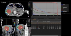
Graph and video demonstrating the full irradiation plan using proton therapy:

The prescription dose is 70.2 Gy (RBE) in fractions of 4.7 Gy (RBE).

Left upper panel displays the axial, left lower the coronal (frontal) and right lower the saggital (side-) view.
Right upper panel dose/volume histograms (DVH´s), meaning the graphic distribution of dose delivered to the volume of specific organs (liver, right kidney, ribs).

DVH shows that only 50% of uninvolved liver tissue received 3.4Gy (RBE); 30%=22.3 Gy (RBE). The average dose to the liver was 18.5 Gy (RBE).

The left kidney was completely spared of any significant radiation dose.

Right kidney reveived doses that should not result in significant functional impairment.: D average= 8.7 Gy (RBE); 30% of Volume received 4.4 Gy (RBE) and 50%=0.5 Gy (RBE)

Ribs that were in the passage of the proton beams received an average dose of 21.21 Gy (RBE)

Colon received minimal dose (D average 0.3 Gy (RBE), in D1%= 6.2 Gy (RBE)

Treatment plan hepatocellular carcinoma

Treatment plan hepatocellular carcinoma (click to enlarge image)

Animation: treatment plan hepatocellular carcinoma

Animation: treatment plan hepatocellular carcinoma (click to start and enlarge animation)

It is of clinical significance that the liver outside the tumor received overall relatively little dose in order to maintain its physiologic function. Its function can be determined and followed in series of blood tests. Specifically in cases of pre-existing liver cirrhosis is the sparing of liver parenchyma of paramount importance. The kidney on the same side receives a dose in the order that will make dysfunction very unlikely. The contralateral kidney receieves no dose. Given sufficiently high dose, ribs have the risks of sustaining fracture. In this case the relatively low dose made a fracture rather unlikely. Note: Spinal cord, spleen, small bowel, stomach, and heart were spared from any meaningful dose or dose at all. Even most modern photon radiation modalities like VMAT or Cyberknife a.o. would have deposited some radiation dose in the treatment process.

+
Example: Pancreas Carcinoma

58-year-old gentleman with poorly differentiated Adenocarcinoma of pancreas head (local stage “borderline resectable”). He underwent chemotherapy. Tumor progression under chemotherapy.

Proton Treatment to a total dose of 37.5 Gy (RBE) (hypofractionated).

Example upper abdomen: pancreas carcinoma

Pancreas carcinoma (click to enlarge image)

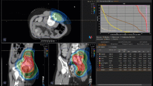
Planning comparison between modern Photon Therapy (VMAT) versus Proton Therapy at MedAustron:

Left upper panel proton plan, left lower panel photon plan.
Right upper panel dose/volume histogram, (DVH) meaning the graphic distribution of dose delivered to the volume of specific organs
Right lower panel the „differential dose“ – meaning the additional dose delivered to normal tissues and structures by photons in comparison to protons. This dose does not contribute to tumor dose.

Treatment plan comparison pancreas carcinoma

Treatment plan comparison pancreas carcinoma (click to enlarge image)

Animation: treatment plan comparison pancreas carcinoma

Animation: treatment plan comparison pancreas carcinoma (click to start and enlarge image)

Note: Of clinical importance is the unnecessary dose given to the surrounding helthy organs/tissues.
The duodenum in particular receives 6 Gy more in VMAT and is in the vicinity of the tumor. The kidneys on both sides receive significantly more dose although these organs are not affected by tumor. The risk of inducing reduced kidney function or even failure is significantly higher than with a proton plan. The spinal cord, the liver and the small intestines receive unnecessary dose that can be avoided by use of protons. The spleen, an organ that is very sensitive to radiation receives in the Photon Plan an average dose of 4.5 Gy (RBE) and using protons less then 2 Gy (RBE) to a very small volume. The risk of developing radiation-induced malignancy later in life is also reduced by the lower radiation exposure of surrounding healthy tissue.

+
FAQ

a. Can particle therapy be combined with systemic therapy?
i. Yes, in pancreatic cancer particle therapy can be combined sequentially with aggressive chemotherapy schema (such as FOLFIRONOX or GEM-ABRAXANE) and can be combined concomitantly with less aggressive regimes such as Gemcitabine or capecitabine. In liver cancer it is preferable to combine particle therapy and chemotherapy in sequence avoiding concomitant use.

b. Can particle therapy be used after surgery?
i. In principle yes and it is routinely done in other tumors (for example skull base tumors). However at present in MedAustron, we are not using particle therapy as post-operative treatment to sterilize the tumor bed in any gastrointestinal tumor.

c. Are there limits regarding the maximum size of treatable tumors?
i. Potentially any size of gastrointestinal tumor can be irradiated. The technical limitation of 20 cm maximum diameter is basically never reached in gastrointestinal tumors.

d. Can particle Therapy be an alternative to surgery?
i. Particle therapy can be an alternative to mutilating surgery (e.g. limb amputation or total sacrectomy or resection of the eye and orbit content). This concept applies in other body districts where mutilation surgical procedure are still compatible with life. For gastrointestinal tumors however mutilating procedures are not possible (for example total liver resection or resection of mesenteric artery and total intestinal resection are not compatible with life). In gastrointestinal tumors surgery, when feasible is the treatment of choice. Particle therapy is used for non resectable cases.

e. Can radiotherapy cause side effects?
i. Yes it can cause side effects and when used at high dose to treat gastrointestinal tumors, they can also be severe or life threatening. The side effects are in general less than those that would be caused by photons radiotherapy if the same high dose were applied.

f. Are there age limits to receive particle therapy?
i. No, age does not play a role for particle therapy.

g. Is there better chance of cure when treated with particles compared to photons?
i. Proton therapy can be used with the same dose as photons radiotherapy. In this case, there is a reduction in risk of toxicity without any increase in the chance of cure.

If a higher dose is applied or if the higher biological efficacy of carbon ions is used, this can lead to an increase in the chance of cure.

+
Literature

1. Rutenberg MS, Nichols RC. Proton beam radiotherapy for pancreas cancer. J Gastrointest Oncol. 2020 Feb;11(1):166-175. doi: 10.21037/jgo.2019.03.02. PMID: 32175120; PMCID: PMC7052755.

2. Kawashiro S, Yamada S, Okamoto M, Ohno T, Nakano T, Shinoto M, Shioyama Y, Nemoto K, Isozaki Y, Tsuji H, Kamada T. Multi-institutional Study of Carbon-ion Radiotherapy for Locally Advanced Pancreatic Cancer: Japan Carbon-ion Radiation Oncology Study Group (J-CROS) Study 1403 Pancreas. Int J Radiat Oncol Biol Phys. 2018 Aug 1;101(5):1212-1221. doi: 10.1016/j.ijrobp.2018.04.057. Epub 2018 May 1. PMID: 29907490

3. Shinoto M, Terashima K, Suefuji H, Matsunobu A, Toyama S, Fukunishi K, Shioyama Y. A single institutional experience of combined carbon-ion radiotherapy and chemotherapy for unresectable locally advanced pancreatic cancer. Radiother Oncol. 2018 Nov;129(2):333-339. doi: 10.1016/j.radonc.2018.08.026. Epub 2018 Sep 14. PMID: 30224179.

4. Mizumoto T, Terashima K, Matsuo Y, Nagano F, Demizu Y, Mima M, Sulaiman NS, Tokumaru S, Okimoto T, Toyama H, Fukumoto T. Proton Radiotherapy for Isolated Local Recurrence of Primary Resected Pancreatic Ductal Adenocarcinoma. Ann Surg Oncol. 2019 Aug;26(8):2587-2594. doi: 10.1245/s10434-019-07471-z. Epub 2019 May 30. PMID: 31147994.

5. Boimel PJ, Berman AT, Li J, Apisarnthanarax S, Both S, Lelionis K, Larson GL, Teitelbaum U, Lukens JN, Ben-Josef E, Metz JM, Plastaras JP. Proton beam reirradiation for locally recurrent pancreatic adenocarcinoma. J Gastrointest Oncol. 2017 Aug;8(4):665-674. doi: 10.21037/jgo.2017.03.04. PMID: 28890817; PMCID: PMC5582048.

6. Liermann J, Ben-Josef E, Syed M, Debus J, Herfarth K, Naumann P. Carbon ion radiotherapy as definitive treatment in locally recurrent pancreatic cancer. Strahlenther Onkol. 2021 Aug 5. doi: 10.1007/s00066-021-01827-9. Epub ahead of print. PMID: 34351449.

7. Kawashiro S, Yamada S, Isozaki Y, Nemoto K, Tsuji H, Kamada T. Carbon-ion radiotherapy for locoregional recurrence after primary surgery for pancreatic cancer. Radiother Oncol. 2018 Oct;129(1):101-104. doi: 10.1016/j.radonc.2018.02.003. Epub 2018 Feb 17. PMID: 29463433.

8. Chuong M, Kaiser A, Molitoris J, Mendez Romero A, Apisarnthanarax S. Proton beam therapy for liver cancers. J Gastrointest Oncol. 2020 Feb;11(1):157-165. doi: 10.21037/jgo.2019.04.02. PMID: 32175119; PMCID: PMC7052772.

9. Kim TH, Koh YH, Kim BH, Kim MJ, Lee JH, Park B, Park JW. Proton beam radiotherapy vs. radiofrequency ablation for recurrent hepatocellular carcinoma: A randomized phase III trial. J Hepatol. 2021 Mar;74(3):603-612. doi: 10.1016/j.jhep.2020.09.026. Epub 2020 Oct 5. PMID: 33031846.

10. Bush DA, Smith JC, Slater JD, Volk ML, Reeves ME, Cheng J, Grove R, de Vera ME. Randomized Clinical Trial Comparing Proton Beam Radiation Therapy with Transarterial Chemoembolization for Hepatocellular Carcinoma: Results of an Interim Analysis. Int J Radiat Oncol Biol Phys. 2016 May 1;95(1):477-482. doi: 10.1016/j.ijrobp.2016.02.027. Epub 2016 Feb 13. PMID: 27084661.

5 — 8

Pelvis

Particle therapy, both proton therapy (PT) and carbon ion radiotherapy (CIRT), is routinely used for various specific pelvic tumors.

In terms of a definitive treatment with curative intent, this includes for example rectal tumors such as tumors of the rectum or anal canal and prostate tumors. In this context of established radiotherapy, conventional irradiation treatment can be replaced by particle therapy. The main motivation is better dose distribution with improved sparing of surrounding healthy organs in both forms of particle therapy offered at MedAustron compared to conventional radiotherapy based on high-energy X-rays, resulting in a substantial reduction of radiotherapy-related toxicity. The established combination of radiotherapy and chemotherapy, as in the case of tumors of the rectum or anal canal and selective gynecological tumors, can also be carried out with proton therapy. This means that conventional photon radiotherapy can be replaced by proton therapy in tumor, but the validated principles of the overall treatment are used.

However, particle therapy can also be used for recurrences of the above-mentioned tumors or other tumors in the pelvis and for radio-resistant or radio-persistent tumors after previous radiation treatment as salvage radiation therapy with curative intent or re-irradiation [see bibliography 2, 3 and 4]. In these scenarios, particle therapy is often the only remaining treatment with curative intent, because conventional radiotherapy or surgical salvage is usually no longer possible due to the pre-existing disease. In these applications of particle therapy in the pelvic area, interdisciplinary discussion with specialist colleagues and experts from other disciplines is very important, for example, in order to insert a protective pre-operatively inserted spacer to distance from strongly pre-stressed organs and to spare them even better during the re-irradiation with particle therapy that is necessary again.

It is not only in particle therapy of pelvic tumors that sparing of healthy surrounding organs is one of the highest priorities in addition to the application of a higher or more effective dose to the tumor or recurrent tumor compared to conventional radiotherapy. Carbon ion radiotherapy in particular is many times more effective in tumor cells than conventional radiotherapy at a given physical dose, and this is virtually independent of the mutation status, oxygen supply to the tumor or higher tumor grading.

+
Indications treated at MedAustron

Indications for definitive particle therapy with curative intent for pelvic tumors are for example:

Proton therapy for not resectable locally (cT3, cT4) and/or logoregionally (with pelvic lymph node metastases, cN+) advanced rectal cancer but without distant metastases (cM0), in combination with established chemotherapy/systemic therapy. In case of distant metastases in primary staging (CT or FDG-PET-CT), interdisciplinary decision with the treating oncologists or surgeons is useful.

Proton therapy for not resectable locally (cT3, cT4) and/or logoregionally (with pelvic lymph node metastases, cN+) advanced anal carcinoma, but without distant metastases (cM0), in combination with established chemotherapy/systemic therapy.

Proton therapy for not resectable locally and/or logoregionally (with pelvic lymph node metastases, cN+) advanced gynecological tumor but without distant metastases (cM0), in combination with established chemotherapy/systemic therapy and/or brachytherapy in a specialized center. Especially in case of metastasis of the pelvic lymph nodes, the advantage of proton therapy over conventional irradiation is greater (better sparing of small intestine, colon, rectum, bladder and sacral nerves).

Proton therapy for low-risk prostate carcinoma or intermediate-risk prostate carcinoma in the context of a study, especially if radical surgery cannot be performed due to anatomy, previous operations or too high anesthetic risk.

Carbon ion radiotherapy for high-risk prostate carcinoma (iPSA > 20 ng/ml or cT2c or cT3 in mpMRI/PSMA-PET or Gleason score 8 to 10) with/without PSMA-avid limited metastasis especially in pelvic lymph nodes combined with hormone deprivation therapy.

Indications for salvage particle therapy in case of non-operability:

Carbon ion radiotherapy as salvage radiation therapy for non-resectable FDG-avid pelvic wall or pelvic recurrences in all of the above tumors but also in other tumors without detection of FDG-avid lesions outside the pelvis in recent re-staging. If necessary, interdisciplinary coordination regarding non-operability and sequential systemic therapy is useful/necessary, if available.

Carbon ion radiotherapy as salvage radiation therapy for PSMA-avid local recurrence of prostate carcinoma after radical prostatectomy without detection of multiple PSMA-avid distant metastases in recent re-staging.

Indications for salvage particle therapy as re-irradiation:

Carbon ion radiotherapy as re-irradiation for FDG-avid pelvic wall recurrences of gynecological tumors after pre-irradiation but with locally controlled primary tumor locally controlled by primary treatment. If necessary, interdisciplinary coordination regarding sequential systemic therapy is useful/necessary, if available.

Carbon ion radiotherapy as re-irradiation for FDG-avid pelvic wall recurrence of colorectal tumors after pre-irradiation but with primary tumor locally controlled by primary treatment. If necessary, interdisciplinary coordination regarding sequential systemic therapy is useful/necessary, if available.

Carbon ion radiotherapy as re-irradiation for FDG-avid pelvic wall recurrences of bladder tumors after pre-irradiation but with primary tumor locally controlled by primary treatment. If necessary, interdisciplinary coordination regarding sequential systemic therapy is useful/necessary, if available.

Carbon ion radiotherapy as re-irradiation for PSMA-avid local recurrence of prostate carcinoma and/or lymph node recurrences of prostate carcinoma after pre-irradiation, but without detection of multiple PSMA-avid distant metastases in recent re-staging.

+
Example: Vaginal Cancer

74-year-old female with a vaginal cancer. The biopsy revealed a squamous cell carcinoma p16 negative, G2. The performed staging detected a 2.4 cm large tumor in introitus vaginae (MRI) cT2 and excluded regional / distant metastatic disease, cN0 cM0.

A normo-fractionated proton therapy to a total dose of 45 Gy (RBE) was performed in 25 fractions. This was fallowed by a PDR brachytherapy boost to EQD2 80 Gy.

Example Indication Pelvis: Vaginal Cancer Plan Comparison

Vaginal cancer plan comparison (click to enlarge image)

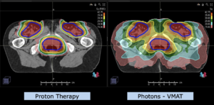
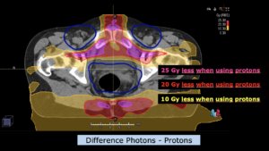
Planning Comparison between modern Photon Therapy (VMAT) with Proton Therapy at MedAustron:

The prescription dose was 45 Gy (RBE) in fractions of 1.8 Gy (RBE).

The images show a comparison between the applied proton therapy and a conventional photon-based VMAT.

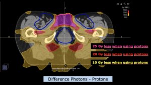
This is a „differential dose“ – meaning the additional dose delivered to normal tissues and structures by photons in comparison to proton therapy, that does not contribute to tumor dose.

Up to 25 Gy (RBE) are delivered unnecessary with photons and are spared by protons e.g. to the external genitals, bladder and soft tissues.

Vaginal cancer dose difference

Vaginal cancer dose difference (click to enlarge image)

Vaginal cancer dose difference II

Vaginal cancer dose difference II (click to enlarge image)

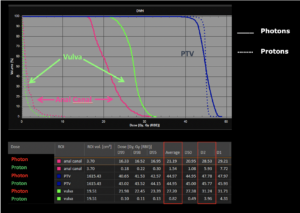
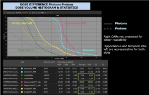
The next image displays dose/volume histograms (DVH‘s), meaning the graphic distribution of dose delivered to the volume of specific organs: vulva in yellow and anal canal in lila (continuous lines correspond to photons and dotted lines to protons).

Huge differences become evident:

Vulva
27.2 Gy (RBE) with photons versus 0.82 Gy (RBE) with protons

Anal canal
21.19 Gy (RBE) with photons versus 1.54 Gy (RBE) with protons

Vaginal cancer dose difference III

Vaginal cancer dose difference III (click to enlarge image)

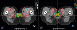
In the video below, the comparison between the applied proton therapy and a conventional photon-based VMAT is shown in the form of the complete treatment plan.

The left panel displays the axial view of the used proton therapy, and the right panel shows the axial view of conventional photon based VMAT technique.

Animation: plan comparison vaginal cancer

Animation: plan comparison vaginal cancer (click to start and enlarge animation)

+
Example: Prostate Cancer

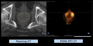
55-year-old patient with a high risk prostate cancer. The biopsy revealed an adenocarcinoma with a Gleason score 4+3=7. The staging was performed with multiparametric MRT & PSMA-PET-CT and detected a multifocal bilateral tumor cT2c and excluded regional / distant metastatic disease, cN0 cM0.

A hypofractionated carbon ion radiotherapy to a total dose of 57.6 Gy (RBE) was performed in 12 fractions.

Example indication pelvis: Prostate cancer

Prostate cancer (click to enlarge image)

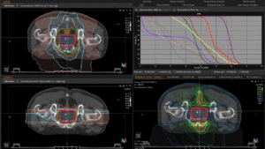
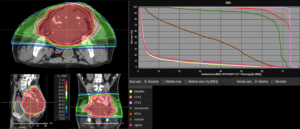
Planning Comparison between modern Photon Therapy (VMAT) with Carbon Ion Radiotherapy at MedAustron:

The prescription dose was 57.6 Gy (RBE) in fractions of 4.8 Gy (RBE).

In the video below, a comparison between the applied carbon ion radiotherapy and a conventional photon-based VMAT is shown. The left upper panel displays the axial view of conventional photon based VMAT technique, and the left lower the axial view of the used carbon ion radiotherapy.

The right lower panel shows the „differential dose“ – meaning the additional dose delivered to normal tissues and structures by photons in comparison to carbon ions up to 35 Gy (RBE), that does not contribute to tumor dose.

Right upper panel displays dose/volume histograms (DVH‘s), meaning the graphic distribution of dose delivered to the volume of specific organs: bladder in yellow, rectum in brown, sigma in green and bowel in purple (continuous lines correspond to photons and dotted lines to carbon ions).

All important organs received significantly less dose with the carbon ion radiotherapy:
Bladder – by 59% less dose (mean dose)
Rectum – by 64% less dose (mean dose)
Bowell – by 39% less dose (in maximal dose)
Colon – by 35% less dose (mean dose) and directly above the rectum (Sigma) by 90% less dose.

Animation: plan comparison prostate cancer

Animation: plan comparison prostate cancer (click to start and enlarge animation)

+
Side Effects

In general, there are no side effects specific to particle therapy, but side effects may occur, as with conventional photon therapy, too. Since less dose reaches the surrounding normal tissue due to the physical properties described, these side effects can be significantly less pronounced.

The tissues or organs in the pelvis surrounding the tumor and not affected by it (e.g. small intestine, colon, rectum, anal canal, bladder, sacral nerves, hip joints, vagina, external genital organs, bulbus penis, etc.) can react very sensitively to ionizing irradiation (radiotherapy) and thus be damaged in the long term. One of the main causes is damage to the smallest blood vessels/capillaries, which in turn leads in the long term to a reduction in blood circulation and corresponding damage to the tissue. These effects can also be observed in areas that received only a low dose. Another cause may be fibrosis, which starts with a short latency after radiotherapy, but is dose-dependent.

Clinically, this can lead to serious acute or chronic side effects. In the case of irradiation of pelvic tumors with high doses, bone and soft tissue necrosis, damage to the sacral nerves, bladder necrosis, perforation and fistulae, as well as intestinal necrosis, perforation and fistulae may be particularly relevant.

All of these effects can occur years after radiotherapy and affect health-related quality of life. The risk of these late effects can usually be significantly reduced with both proton and carbon ion radiotherapy due to the substantial minimization of the dose outside the target volume/tumor in the surrounding healthy organs and tissues compared to conventional irradiation.

For comparison, long-term data from conventional photon radiotherapy in the randomized study ASCENDE-RT (“Androgen Suppression Combined with Elective Nodal and Dose Escalated Radiation Therapy”) for dose-escalated and intensity-modulated photon radiotherapy of prostate carcinomas with total doses of 78 Gy (see bibliography 5) show high-grade chronic toxicities of bladder tumors (5.2%) and intestine (gastrointestinal) tumors) (3.2%) (see bibliography 6).

Compared to these data, Japanese long-term data with carbon ion radiotherapy show no high-grade chronic toxicities of bladder tumors (0.3%) and gastrointestinal tumors (0%) (see bibliography 1).

+
FAQ

a. Can particle therapy be combined with systemic therapy?
i. Depending on the tumor, particle therapy can be combined with systemic therapy, as it is with conventional radiotherapy as well. In such cases, the indication is made on an interdisciplinary basis (medical oncologist and radiation oncologist). See also the explanations for the individual indications in section II.

b. Is particle therapy possible after surgery as well?
i. Particle therapy can also be performed after surgery, especially if the surgeon could not remove everything, it is also indicated. This is especially the case with pelvic lymph node metastases, also because micro-metastases outside the surgical area should be expected. In such cases, coincident irradiation of an entire anatomical region would be useful.

c. Is there a limitation on the tumor size in particle therapy?
i. In principle, tumors of any size can be treated with particle therapy.

d. Can particle therapy replace surgery?
i. If a tumor is accessible to surgery, this is the treatment of choice in the majority of cases. In some situations, surgery is associated with high risk, so that irradiation can be performed as an option with curative intent. However, a definitive treatment with curative intent is established for example for locally or locoregionally advanced rectal tumors such as tumors of the anal canal, gynecological tumors and prostate tumors.

e. Can particle therapy cause side effects?
i. Locally, similar to conventional irradiation, reactions can also occur in the surrounding normal tissue. As a rule, however, these are less pronounced, since due to the superior physical properties of particle therapy, these surrounding tissues or organs are exposed to a significantly lower dose.

f. Are the chances of cure higher after particle therapy than after conventional irradiation?
i. The biological effect of proton therapy is very similar to that of photon therapy. Thus, if proton therapy were applied at the same dose, the chances of cure would be comparable after proton therapy as after conventional irradiation. However, due to the lower normal tissue exposure in special situations, the total dose is significantly increased with proton therapy and consequently an improvement in local tumor control is highly likely to be achieved.

Nevertheless, the biological effect of carbon ion radiotherapy is significantly higher compared to photon therapy but also compared to proton therapy. Therefore, carbon ion radiotherapy should generally be used with higher priority in radiation-resistant tumors or in the sense of re-irradiation in radiation-recurrent tumors after pre-irradiation.

g. Are there age limits for particle therapy?
i. In principle, patients of any age who are indicated for radiotherapy can be treated with both forms of particle therapy at MedAustron. However, the general condition of the affected patients should be sufficiently preserved to make such treatment appear reasonable.

+
Literature

1. Kawamura H, Kubo N, Sato H, et al. Moderately hypofractionated carbon ion radiotherapy for prostate cancer; a prospective observational study “GUNMA0702” BMC Cancer. 2020; 20: 75.

2. Habermehl D, Wagner M, Ellerbrock M, Büchler MW, Jäkel O et al. Reirradiation Using Carbon Ions in Patients with Locally Recurrent Rectal Cancer at HIT: First Results. Ann Surg Oncol. 2015; 22(6): 2068-74.

3. Yamada S, Kamada T, Ebner DK, Shinoto M, Terashima K et al. Carbon-Ion
Radiation Therapy for Pelvic Recurrence of Rectal Cancer. Int J Radiat Oncol Biol
Phys. 2016 Sep 1; 96(1): 93-101.

4. Berman AT, Both S, Sharkoski T, Goldrath K, Tochner Z. Proton Reirradiation of Recurrent Rectal Cancer: Dosimetric Comparison, Toxicities, and Preliminary Outcomes. International Journal of Particle Therapy. 2014; 1(1): 2-13

5. Morris WJ, Tyldesley S, Rodda S, et al. Androgen Suppression Combined with Elective Nodal and Dose Escalated Radiation Therapy (the ASCENDE-RT Trial): An Analysis of Survival Endpoints for a Randomized Trial Comparing a Low-Dose-Rate Brachytherapy Boost with Dose-Escalated External Beam Boost for High- and Intermediate-risk Prostate Cancer. Int J Radiat Oncol Biol Phys. 2017; 98(2): 275-285.

6. Rodda S, Tyldesley S, Morris WJ, et al. ASCENDE-RT: An Analysis of Treatment Related Morbidity for a Randomized Trial Comparing a Low-Dose-Rate Brachytherapy Boost with Dose-Escalated External Beam Boost for High- and Intermediate-risl Prostate Cancer. Int J Radiat Oncol Biol Phys. 2017; 98(2): 286-295.

6 — 8

Sarcoma

Particle therapy with both protons and carbon ions has been used since more than 30 years in the treatment of sarcoma.

Both particles can achieve an optimal sparing of the noninvolved healthy tissues and therefore reduce unwanted side effects and toxicity. Because of this advantage in dose distribution, both particles allow safe escalation of the dose. Moreover, carbon ions, thanks to their radiobiological properties, can overcome the radio resistance of tumors (such as chondrosarcoma and osteosarcoma) that are difficult to control with other kinds of radiotherapy.

+
Tumors

With the term sarcoma, we designate a large variety of tumors originating from the connective tissue that differ significantly in all relevant aspects. According to the World Health Organization classification, there are more than 20 different groups of sarcomas and more than 150 different subtypes. Given this complexity, it is exceedingly difficult to make general statements and an evaluation of the individual case is always mandatory.

There are however some general considerations that are valid for the most common scenarios.

Tumors of the limbs (i.e. arms and legs) can be successfully treated, in the majority of cases, also without particle therapy. More specifically osteosarcomas of the limbs are treated by a combination of chemotherapy and conservative surgery and soft tissue sarcoma of the limbs are treated by a combination of photons radiotherapy and conservative surgery. For those tumors the role of particle therapy is minimal. Single cases that would not be candidate to conservative surgery can be evaluated to explore the possibility of avoiding limb amputation. In addition, particle therapy can, in selective cases, avoid radiation dose to uninvolved muscle compartments, thereby improving functional outcome.

Gastrointestinal Stromal Tumors have a well-established treatment protocol based on a combination of surgery and targeted systemic therapy.

Particle therapy can play a role in tumors originating in the head and neck, thorax, abdomen and pelvis. Particle therapy is used to increase the local control of the primary tumor in non-metastatic patients. In this patients group, the increase in local control can correspond to an increase in the probability of cure and survival. Generally, particle therapy is not used in already metastatic patients, it can however be evaluated in selected cases (either if the metastasis have an indolent growth that does not determine the prognosis in the mid-term and/or if there is no other way to achieve symptoms control).

Particle therapy can be used in combination with surgery or as unique local treatment modality. According to the histology, systemic therapy can be added to the treatment.

+
Indications treated at MedAustron

Vertebral and Sacral Chordoma

Particle therapy (alone or in combination with surgery) is the gold standard for the treatment of chordoma. Systemic therapy plays a minor role and is used only in metastatic patient or in very advanced local relapses not amenable to further local therapies.

Sacral chordoma patients should be evaluated by an expert surgeon and by an expert radiation oncologist. According to the level of invasion of the tumor (upper sacrum S1-S2 versus lower sacrum S3-S5) the sequelae of surgery may be more or less invalidating. The therapy of choice can be surgery followed by proton therapy or radical carbon ion radiotherapy. If the tumor abuts or compresses the rectum and/or sigmoid colon, a preliminary surgical procedure may be necessary in order to insert a spacer between tumor and intestine.

For tumors arising in the mobile spine, the two most critical factors are the relationship between tumor and spinal cord and the relationship between tumor and vertebral arteries.

In the cervical spine, the presence of both spinal cord and vertebral arteries makes a radical resection extremely problematic. The optimal treatment typically consists of surgery followed by particle therapy. If the tumor abuts the spinal cord, even particle therapy becomes problematic. A minimum distance is in fact needed between the target that must be irradiated with a very high dose and some radiosensitive critical organs. In these cases, a so-called “separating surgery” should be considered to decompress the spinal cord resecting the component of the tumor inside the spinal canal. This kind of surgery (sometimes also called intra-lesional debulking) is on the one hand much less invasive than a complete tumor resection but on the other hand, it is by itself not adequate and cannot control the tumor, its sole purpose is to enable safe delivery of high dose particle radiotherapy. The choice between protons and carbon ions will mainly be driven by the amount of macroscopic residual disease.

In the thoracic spine and in the upper lumbar spine the vertebral arteries do not limit the possibility of complete surgical resection. In this district, the best therapy is usually a gross macroscopic surgical resection followed by postoperative proton therapy. The spinal cord does not extend to the lower lumbar spine (L3-L5); in this district the spinal canal content is limited to the (more radio-resistant) cauda equina and therefore radical particle therapy (preferably with carbon ions) is often possible even without surgery.

In selected cases, a sandwich schedule with preoperative particle therapy followed by surgery and finally by postoperative particle therapy may be needed. This approach is usually selected in bulky tumor that need both surgery and radiation but in which the resection procedure is at risk of seeding the tumor to other noninvolved tissues.

It is mandatory to discuss the overall treatment strategy upfront. Special care is needed in selecting the kind of spinal stabilization devices and of reconstruction for the resected vertebral bodies in order to minimize the issues related with imaging artefacts and particle range.

Inoperable osteosarcoma

The established treatment of osteosarcoma is preoperative chemotherapy followed by radical surgery and post-operative chemotherapy. Radical surgery is hardly ever feasible in osteosarcoma of the cervical spine and skull base. Osteosarcoma of the pelvis, thoracic wall, maxillary region, thoraco-lumbar spine and sacrum should be evaluated by orthopedic surgeons with a specific expertise and in a referral center. High dose carbon ion radiotherapy is used as radical local therapy, in alternative to surgery, for cases that are not technically resectable (e.g. an osteosarcoma of the cervical spine encasing both vertebral arteries) or in medically inoperable patients (due to age and comorbidities) or in patients that refuse a mutilating surgical procedure (e.g. a total sacrectomy with high risk of double incontinence or an hemipelvectomy with limb amputation).

Chemotherapy will be given before and after carbon ion radiotherapy (as it would have been done if the patient had been operated).

Sometimes specific surgical procedures may be necessary before high dose carbon ion radiotherapy.

Separating surgery may be needed to decompress the spinal cord (see the paragraph on chordoma above)
In some cases, it might be necessary to surgically insert a spacer to separate the bowel loops from the area that must be treated with a high dose (see the paragraph on chordoma above).

The dose needed to control osteosarcoma can be toxic for the surrounding noninvolved tissues. It is sometimes necessary to perform surgical procedures to treat the sequelae of high dose carbon ions radiotherapy. For example, surgical spinal stabilization might be necessary to prevent or to treat vertebral body fracture after radical CIRT for lumbar spine osteosarcoma or surgical maxillectomy with prosthetic reconstruction might be necessary to treat osteoradionecrosis induced by CIRT for maxillary osteosarcoma.

Chondrosarcoma

Systemic therapy plays almost no role in the treatment of chondrosarcoma. For chondrosarcomas of the extremities surgery, when possible, is the treatment of choice for many patients. Post-operative radiotherapy is typically not indicated after a radical resection with clean margins. Particle therapy can be used in those cases that are not technically resectable or for those patients that are not medically operable or refuse a mutilating surgical procedure. As already discussed in the previous paragraphs, preliminary surgical procedures (such as spinal cord decompression or intestinal spacer) may be necessary before particle therapy.

Ewing sarcoma

Ewing sarcomas typically originate in the bones; this tumor responds well to chemotherapy, which is the initial treatment of choice. According to the site of origin, local extension and degree of response to chemotherapy, the local treatment can be either surgery + radiotherapy or radiotherapy alone.

The doses required to control these tumors are lower than those necessary for the more radio-resistant histologies described above (typically less than 60 Gy in conventional fractionation).

Ewing sarcoma is typically a tumor of the young age. A long survival and cure is often possible. In Ewing sarcoma particle therapy is used (post operatively or exclusively) mainly to reduce the integral dose and the related unwanted long term side effects. Most patients are treated with proton therapy and CIRT is reserved to those few cases progressing under chemotherapy.

Rhabdomyosarcoma

This tumor is typical in the pediatric age (see section on pediatric tumors). The initial treatment consists in systemic chemotherapy.

The choice of local therapy depends on several factors and in the most favorable cases radiotherapy can be altogether avoided. If radiotherapy is needed proton therapy is the treatment of choice. The doses needed to control rhabdomyosarcomas are moderate (< 60 Gy) and therefore the main focus is reducing the integral dose and the related unwanted long term side effects.

Retroperitoneal sarcoma

Radical surgery is the treatment of choice for retroperitoneal sarcoma. Preoperative RT has been tested in randomized trial and has been shown to be of little value for those histologies (leiomyosarcoma and undifferentiated pleomorphic sarcoma) in which the relapse pattern is dominated by distant metastasis. Preoperative radiotherapy is on the contrary recommended in retroperitoneal liposarcoma. Proton therapy is used in preoperative treatment of liposarcoma to reduce integral dose and toxicity. Carbon ion radiotherapy can be used in selected cases that are not candidate to radical surgery.

Head and neck sarcoma

Due to the anatomical complexity and the presence of many non-sacrificable structure radical surgery is rarely feasible in this district. When radical surgery (followed by surgical reconstruction) is feasible (e.g. in osteosarcoma of the mandible) it should be considered the first option.

In the majority of head and neck sarcoma that are not candidate to radical surgery, particle therapy can be used as an alternative to surgery. Reconstructive surgery may be needed to treat the sequelae of high dose particle therapy.

Skull Base Sarcomas

The skull base is a distinctly important anatomic structure that separates facial structures, mouth and pharynx from the brain. It is not just a bony separation but also allows important nerves and blood vessels to connect between the rest of the body and the brain. See also separate section on Skull Base Tumors.

In general, sarcomas require radiation dosages for complete tumor cell kill that exceed normal tissue tolerances of almost all organs in the body. Therefore, sarcomas arising in the skull base are typically in proximity or might even compress important and vital structures and organs, for example, the brain stem, optic nerves, optic chiasm and others.

Sarcomas of the skull base were one of the first indications treated with protons more than 30 years ago that have proven the superiority of particle over conventional radiation therapy. Protons are very suitable. In addition, carbon ion therapy is now routinely used since many of the sarcomas are relatively radio resistant in nature and carbon ion therapy can overcome the radio-resistance.

Paraspinal Sarcomas

Most of the sarcomas already mentioned can occur along the axial skeleton and sacrum in immediate proximity to the spinal cord. Examples are chordomas, chondrosarcomas, osteogenic sarcomas, Ewing’s sarcomas, malignant peripheral nerve sheath tumors, but also in principle benign but locally aggressive tumors like chondroblastomas and osteoblastomas. Frequently they cannot be completely resected without sacrificing nerve roots or endangering function of the spinal cord or in the neck region without endangering functioning of the vertebral artery (in case it cannot be occluded).

Not only does a subtotal resection pose a significant challenge to the radiation oncologist, matters are often made even more difficult by the fact that the surgeon at the time of operation had to resect portions of vertebral bodies that render the situation unstable and the surgeon therefore had to insert rods tightened with screws to stabilize the skeleton. These patients are reviewed on an individual basis for suitability for particle therapy. Often they are candidates for particle therapy up front, i.e. before planned surgery. An interdisciplinary approach, i.e. discussions with the surgeons, is of critical importance to provide optimum individualized management.

Other histologies

For other soft tissue sarcomas arising in the head and neck and in the trunk the decision algorithm can, to a given extend, be extrapolated from soft tissue sarcoma of the limbs.

The three bad prognostic factors are high grade, bulky lesion (> 5 cm) and non-superficial tumors.

When 2 or 3 of these factors are present, the therapy of choice is radical surgery followed by postoperative radiotherapy. Proton therapy can be used to reduce integral dose and spare toxicity. Carbon ion radiotherapy can be used as an alternative for surgery in those cases that are not technically resectable or for those patient that are not medically operable or refuse a mutilating surgical procedure. The role of chemotherapy and the need to tailor chemotherapy schedule according to histology are still a matter of debate.

Treatment of local recurrence

A local recurrence in a non-previously irradiated area and in the absence of distant metastasis should always be evaluated for a second therapy with curative intent. As a general rule the presence of a local relapse creates doubt regarding the microscopic contamination of the initial surgical bed and reduces the theoretical value of a second radical resection. The choice between second surgery or radical particle therapy must be mad on a case by case basis considering the risk of toxicity and probability of success of both approaches.

Re-irradiation

A re-irradiation of a local failure after photons radiotherapy or after a previous particle therapy is always challenging. The feasibility must be evaluated examining in detail the dose distribution of the first treatment. The limiting factor is typically the dose already received by the critical organ. The favorable physical characteristics of particle therapy make it an ideal tool for re-irradiation, there are however cases where a second course of radiotherapy is not possible even with particle therapy (e.g. a relapse compressing the spinal cord in an area that has already received a dose in excess of 50 Gy in the first course of RT).

+
Example: Sacral Chordoma

41-year-old man with sacral chordoma. Complete urinary incontinence and severe pain. The patient refuses mutilating surgery and is referred for carbon ions radiotherapy.

Treated with exclusive carbon ion radiotherapy 73.6 Gy (RBE)

Example: sacral chordoma

Sacral chordoma (click to enlarge image)

Graph and video demonstrating the full radiation plan using CARBON ION radiotherapy:

Left side: Carbon Ion plan in 3 views: axial, sagittal (side) and coronal (frontal) view.
Right side: dose/volume histograms (DVH‘s), meaning the graphic distribution of dose delivered to the volume of specific organs – in this case bladder, rectum, sigmoig colon.

Treatment plan sacral chordoma

Treatment plan sacral chordoma (click to enlarge image)

Animation: treatment plan sacral chroma

Animation: treatment plan sacral chroma (click to start and enlarge animation)

Note: the pelvic organs ( i.e. urinary bladder, rectum and sigmoid colon) are optimally spared. Even the nerve roots in close proximity to the tumor can be selectively spared.
Photon radiotherapy would not be considered a curative treatment for this bulky tumor with radioresistant histology.

+
Example: Sarcoma of the Costo-Vertebral Joint

50-year-old woman with undifferentiated sarcoma of the costo-vertebral joint at the level of the IXth thoracic vertebra. Previous radiotherapy for Hodgkin Lymphoma 25 years ago. Surgical resection was performed but could not achieve microscopic radicality. The patient is referred for post-operative proton therapy.

Treated with proton therapy to 60 Gy (RBE).

Example: sarcoma

Sarcoma (click to enlarge image)

Graph and video demonstrating the full irradiation plan using proton therapy:

Left side: Proton plan in 3 views: axial, sagittal (side) and coronal (frontal) view.
Right side: dose/volume histograms (DVH‘s), meaning the graphic distribution of dose delivered to the volume of specific organs – in this case spinal cord, lungs, esophagus and stomach.

Treatment plan sarcoma

Treatment plan sarcoma (click to enlarge image)

Animation: Treatment plan sarcoma

Animation: Treatment plan sarcoma (click to start and enlarge animation)

Note: the thoracic and abdominal organs ( i.e. lungs, heart, esophagus, stomach and liver ) are almost or completely spared. The dose to spinal cord is limited to a safe dose level.

+
Side Effects

The unwanted side effects of particle therapy are similar to those of conventional photons based radiation therapy. However, the physical properties of particle therapy are more favorable than those of photons and this leads to a better normal tissue sparing. The toxicity of any particle plan is lower than that of the same curative dose delivered with photons RT. It is however also true that some of the indication for particle therapies would not have been treated at all with photons RT with curative intent (e.g. bulky non resected pelvic osteosarcoma). In conclusion, the toxicity of curative particle therapy for sarcoma can be substantial and might even require surgical treatment.

Particle therapy for sarcomas following either a total resection or a subtotal resection with a small amount of residual disease is typically well tolerated with a small risk of severe long term side effects. Any such risks are highly dependent on the individual situation and best discussed with the radiation oncologist. In case of unresectable, large disease particle therapy, in particular also carbon ion therapy, can often still be delivered with true curative intent, i.e. to attempt cure. However, high dosages are required and they might result in a risk of deficit or potential loss of nerve function in this area or possibly in a fracture of a bone already involved and very often already partially destroyed by tumor. Again, these situations are highly individualized and it has to be remembered that any surgery would have required major resections potentially or with the risk of resulting in major loss of function due to surgery.

+
FAQ

a. Can particle therapy be combined with systemic therapy?
i. Yes, it is theoretically possible and in other tumors (e.g. mucosal melanoma or pancreatic cancer) concomitant particle therapy + systemic therapy is routinely done. In sarcoma systemic therapy and particle therapy are often used together but they are normally delivered sequentially rather than concomitantly.

b. Can particle therapy be used after surgery?
i. Yes, either with a lower dose to kill the residual microscopic disease or with high dose in case of residual macroscopic tumor

c. Are there limits regarding the maximum size of treatable tumors?
i. Due to technical reason, it is exceedingly difficult to treat at MedAustron tumors with a maximum diameter larger than 20 cm. These patients are routinely referred to other particle facilities.

d. Can particle therapy be an alternative to surgery?
i. In principle, radical surgery is always the treatment of choice. The only exception is sacral chordoma where radical surgery or radical particle therapy are both considered valid options. In many cases particle therapy can however be used instead of surgery if a radical resection is not technically feasible or if the patient is not medically able to tolerate the procedure or if the patient reuses a mutilating surgical procedure.

e. Can particle therapy cause side effects?
i. Yes, especially when used at high doses in the radical treatment of a non-resected sarcoma particle therapy can also result in severe side effects.

f. Are there age limits to receive particle therapy?
i. No, proton therapy is used to reduce side effects in children and even in infants, elderly patient can be treated without age limits. Patients older than 90 years are routinely treated in MedAustron.

+
Literature

1. Komatsu S, Okamoto M, Shiba S, Kaminuma T, Okazaki S, Kiyohara H, Yanagawa T, Nakano T, Ohno T. Prospective Evaluation of Quality of Life and Functional Outcomes after Carbon Ion Radiotherapy for Inoperable Bone and Soft Tissue Sarcomas. Cancers (Basel). 2021 May 25;13(11):2591. doi: 10.3390/cancers13112591. PMID: 34070569; PMCID: PMC8199366.

2. Shiba S, Okamoto M, Kiyohara H, Okazaki S, Kaminuma T, Shibuya K, Kohama I, Saito K, Yanagawa T, Chikuda H, Nakano T, Ohno T. Impact of Carbon Ion Radiotherapy on Inoperable Bone Sarcoma. Cancers (Basel). 2021 Mar 4;13(5):1099. doi: 10.3390/cancers13051099. PMID: 33806515; PMCID: PMC7961536.

3. Shiba S, Okamoto M, Tashiro M, Ogawa H, Osone K, Yanagawa T, Kohama I, Okazaki S, Miyasaka Y, Osu N, Chikuda H, Saeki H, Ohno T. Rectal dose-sparing effect with bioabsorbable spacer placement in carbon ion radiotherapy for sacral chordoma: dosimetric comparison of a simulation study. J Radiat Res. 2021 May 12;62(3):549-555. doi: 10.1093/jrr/rrab013. PMID: 33783533; PMCID: PMC8127650.

4. Seidensaal K, Kieser M, Hommertgen A, Jaekel C, Harrabi SB, Herfarth K, Mechtesheimer G, Lehner B, Schneider M, Nienhueser H, Fröhling S, Egerer G, Debus J, Uhl M. Neoadjuvant irradiation of retroperitoneal soft tissue sarcoma with ions (Retro-Ion): study protocol for a randomized phase II pilot trial. Trials. 2021 Feb 12;22(1):134. doi: 10.1186/s13063-021-05069-z. PMID: 33579340; PMCID: PMC7881463.

5. Takenaka S, Araki N, Outani H, Hamada KI, Yoshikawa H, Kamada T, Imai R. Complication rate, functional outcomes, and risk factors associated with carbon ion radiotherapy for patients with unresectable pelvic bone sarcoma. Cancer. 2020 Sep 15;126(18):4188-4196. doi: 10.1002/cncr.33082. Epub 2020 Jul 13. PMID: 32658315.

6. Matsumoto Y, Matsunobu A, Kawaguchi K, Hayashida M, Iida K, Saiwai H, Okada S, Endo M, Setsu N, Fujiwara T, Baba S, Nomoto S, Nakashima Y. Clinical results of carbon-ion radiotherapy with separation surgery for primary spine/paraspinal sarcomas. Int J Clin Oncol. 2019 Nov;24(11):1490-1497. doi: 10.1007/s10147-019-01505-y. Epub 2019 Jul 6. PMID: 31280398.

7. Yang J, Gao J, Qiu X, Hu J, Hu W, Wu X, Zhang C, Ji T, Kong L, Lu JJ. Intensity-Modulated Proton and Carbon-Ion Radiation Therapy in the Management of Head and Neck Sarcomas. Cancer Med. 2019 Aug;8(10):4574-4586. doi: 10.1002/cam4.2319. Epub 2019 Jun 23. PMID: 31231939; PMCID: PMC6712452.

8. Hayashi K, Koto M, Ikawa H, Hagiwara Y, Tsuji H, Ogawa K, Kamada T. Feasibility of Re-irradiation using carbon ions for recurrent head and neck malignancies after carbon-ion radiotherapy. Radiother Oncol. 2019 Jul;136:148-153. doi: 10.1016/j.radonc.2019.04.007. Epub 2019 Apr 19. PMID: 31015117.

9. Vitolo V, Barcellini A, Fossati P, Fiore MR, Vischioni B, Iannalfi A, Facoetti A, Bonora M, Ronchi S, D’Ippolito E, Petrucci R, Viselner G, Preda L, Ciocca M, Valvo F, Orecchia R. Carbon Ion Radiotherapy in the Management of Unusual Liposarcomas: A Case Report. In Vivo. 2019 Mar-Apr;33(2):529-533. doi: 10.21873/invivo.11506. PMID: 30804137; PMCID: PMC6506314.

10. Ando K, Kobayashi K, Machino M, Ota K, Morozumi M, Tanaka S, Imai R, Nishida Y, Ishiguro N, Imagama S. Fusion surgery with instrumentation following carbon ion radiotherapy for primary lumbar tumors: A case series. J Clin Neurosci. 2019 Apr;62:264-268. doi: 10.1016/j.jocn.2019.01.001. Epub 2019 Jan 14. PMID: 30655237.

11. Imai R, Kamada T, Araki N; Working Group for Carbon Ion Radiotherapy for Bone and Soft Tissue Sarcomas. Carbon ion radiotherapy for unresectable localized axial soft tissue sarcoma. Cancer Med. 2018 Sep;7(9):4308-4314. doi: 10.1002/cam4.1679. Epub 2018 Jul 20. PMID: 30030906; PMCID: PMC6143931.

12. Yang J, Gao J, Wu X, Hu J, Hu W, Kong L, Lu JJ. Salvage Carbon Ion Radiation Therapy for Locally Recurrent or Radiation-Induced Second Primary Sarcoma of the Head and Neck. J Cancer. 2018 Jun 4;9(12):2215-2223. doi: 10.7150/jca.24313. PMID: 29937942; PMCID: PMC6010679.

13. Imai R, Kamada T, Araki N; WORKING GROUP FOR BONE and SOFT-TISSUE SARCOMAS. Clinical Efficacy of Carbon Ion Radiotherapy for Unresectable Chondrosarcomas. Anticancer Res. 2017 Dec;37(12):6959-6964. doi: 10.21873/anticanres.12162. PMID: 29187480.

14. Matsumoto Y, Shinoto M, Endo M, Setsu N, Iida K, Fukushi JI, Kawaguchi K, Okada S, Bekki H, Imai R, Kamada T, Shioyama Y, Nakashima Y. Evaluation of Risk Factors for Vertebral Compression Fracture after Carbon-Ion Radiotherapy for Primary Spinal and Paraspinal Sarcoma. Biomed Res Int. 2017;2017:9467402. doi: 10.1155/2017/9467402. Epub 2017 Jul 26. PMID: 28815184; PMCID: PMC5549470.

15. Demizu Y, Jin D, Sulaiman NS, Nagano F, Terashima K, Tokumaru S, Akagi T, Fujii O, Daimon T, Sasaki R, Fuwa N, Okimoto T. Particle Therapy Using Protons or Carbon Ions for Unresectable or Incompletely Resected Bone and Soft Tissue Sarcomas of the Pelvis. Int J Radiat Oncol Biol Phys. 2017 Jun 1;98(2):367-374. doi: 10.1016/j.ijrobp.2017.02.030. Epub 2017 Feb 22. PMID: 28463156.

16. Imai R, Kamada T, Araki N; Working Group for Bone and Soft Tissue Sarcomas. Carbon Ion Radiation Therapy for Unresectable Sacral Chordoma: An Analysis of 188 Cases. Int J Radiat Oncol Biol Phys. 2016 May 1;95(1):322-327. doi: 10.1016/j.ijrobp.2016.02.012. Epub 2016 Feb 8. PMID: 27084649.

17. Seidensaal K, Mattke M, Haufe S, Rathke H, Haberkorn U, Bougatf N, Kudak A, Blattmann C, Oertel S, Kirchner M, Buesch C, Kieser M, Herfarth K, Kulozik A, Debus J, Uhl M, Harrabi SB. The role of combined ion-beam radiotherapy (CIBRT) with protons and carbon ions in a multimodal treatment strategy of inoperable osteosarcoma. Radiother Oncol. 2021 Jun;159:8-16. doi: 10.1016/j.radonc.2021.01.029. Epub 2021 Feb 4. PMID: 33549644.

18. Buszek SM, Ludmir EB, Grosshans DR, McAleer MF, McGovern SL, Harrison DJ, Okcu MF, Chintagumpala MM, Mahajan A, Paulino AC. Disease Control and Patterns of Failure After Proton Beam Therapy for Rhabdomyosarcoma. Int J Radiat Oncol Biol Phys. 2021 Mar 1;109(3):718-725. doi: 10.1016/j.ijrobp.2020.09.050. PMID: 33516439.

19. Uezono H, Indelicato DJ, Rotondo RL, Mailhot Vega RB, Bradfield SM, Morris CG, Bradley JA. Treatment Outcomes After Proton Therapy for Ewing Sarcoma of the Pelvis. Int J Radiat Oncol Biol Phys. 2020 Aug 1;107(5):974-981. doi: 10.1016/j.ijrobp.2020.04.043. Epub 2020 May 8. PMID: 32437922.

20. DeLaney TF, Yock TI, Paganetti H. Assessing second cancer risk after primary cancer treatment with photon or proton radiotherapy. Cancer. 2020 Aug 1;126(15):3397-3399. doi: 10.1002/cncr.32936. Epub 2020 May 19. PMID: 32426850.

21. Indelicato DJ, Rotondo RL, Krasin MJ, Mailhot Vega RB, Uezono H, Bradfield S, Agarwal V, Morris CG, Bradley JA. Outcomes Following Proton Therapy for Group III Pelvic Rhabdomyosarcoma. Int J Radiat Oncol Biol Phys. 2020 Apr 1;106(5):968-976. doi: 10.1016/j.ijrobp.2019.12.036. Epub 2020 Jan 25. PMID: 31987977.

22. Stieb S, Snider JW 3rd, Placidi L, Kliebsch U, Lomax AJ, Schneider RA, Weber DC. Long-Term Clinical Safety of High-Dose Proton Radiation Therapy Delivered With Pencil Beam Scanning Technique for Extracranial Chordomas and Chondrosarcomas in Adult Patients: Clinical Evidence of Spinal Cord Tolerance. Int J Radiat Oncol Biol Phys. 2018 Jan 1;100(1):218-225. doi: 10.1016/j.ijrobp.2017.08.037. Epub 2017 Sep 4. PMID: 29029887.

23. Guttmann DM, Frick MA, Carmona R, Deville C Jr, Levin WP, Berman AT, Chinniah C, Hahn SM, Plastaras JP, Simone CB 2nd. A prospective study of proton reirradiation for recurrent and secondary soft tissue sarcoma. Radiother Oncol. 2017 Aug;124(2):271-276. doi: 10.1016/j.radonc.2017.06.024. Epub 2017 Jul 8. PMID: 28697854.

24. Demizu Y, Jin D, Sulaiman NS, Nagano F, Terashima K, Tokumaru S, Akagi T, Fujii O, Daimon T, Sasaki R, Fuwa N, Okimoto T. Particle Therapy Using Protons or Carbon Ions for Unresectable or Incompletely Resected Bone and Soft Tissue Sarcomas of the Pelvis. Int J Radiat Oncol Biol Phys. 2017 Jun 1;98(2):367-374. doi: 10.1016/j.ijrobp.2017.02.030. Epub 2017 Feb 22. PMID: 28463156.

25. Demizu Y, Mizumoto M, Onoe T, Nakamura N, Kikuchi Y, Shibata T, Okimoto T, Sakurai H, Akimoto T, Ono K, Daimon T, Murayama S. Proton beam therapy for bone sarcomas of the skull base and spine: A retrospective nationwide multicenter study in Japan. Cancer Sci. 2017 May;108(5):972-977. doi: 10.1111/cas.13192. Epub 2017 Apr 24. PMID: 28182320; PMCID: PMC5448607.

26. Indelicato DJ, Rotondo RL, Begosh-Mayne D, Scarborough MT, Gibbs CP, Morris CG, Mendenhall WM. A Prospective Outcomes Study of Proton Therapy for Chordomas and Chondrosarcomas of the Spine. Int J Radiat Oncol Biol Phys. 2016 May 1;95(1):297-303. doi: 10.1016/j.ijrobp.2016.01.057. PMID: 27084648.

27. Holliday EB, Mitra HS, Somerson JS, Rhines LD, Mahajan A, Brown PD, Grosshans DR. Postoperative proton therapy for chordomas and chondrosarcomas of the spine: adjuvant versus salvage radiation therapy. Spine (Phila Pa 1976). 2015 Apr 15;40(8):544-9. doi: 10.1097/BRS.0000000000000804. PMID: 25627289.

28. Ciernik IF, Niemierko A, Harmon DC, Kobayashi W, Chen YL, Yock TI, Ebb DH, Choy E, Raskin KA, Liebsch N, Hornicek FJ, Delaney TF. Proton-based radiotherapy for unresectable or incompletely resected osteosarcoma. Cancer. 2011 Oct 1;117(19):4522-30. doi: 10.1002/cncr.26037. Epub 2011 Mar 29. PMID: 21448934; PMCID: PMC3716000.

29. DeLaney TF, Liebsch NJ, Pedlow FX, Adams J, Dean S, Yeap BY, McManus P, Rosenberg AE, Nielsen GP, Harmon DC, Spiro IJ, Raskin KA, Suit HD, Yoon SS, Hornicek FJ. Phase II study of high-dose photon/proton radiotherapy in the management of spine sarcomas. Int J Radiat Oncol Biol Phys. 2009 Jul 1;74(3):732-9. doi: 10.1016/j.ijrobp.2008.08.058. Epub 2008 Dec 25. PMID: 19095372; PMCID: PMC2734911.

7 — 8

Pediatric Malignancies

In the early 90s proton therapy was introduced to the pediatric radiation oncology community after initial radiation therapy planning comparisons demonstrated a clear advantage for CNS tumors and sarcomas.

Treatment planning comparisons, whereby the best plan of either state-of-the-art photon (x-ray) therapy is compared with proton therapy, is an excellent tool to understand in an individual case the possible advantages of proton therapy. With very few exceptions, virtually all proton and photon comparisons demonstrated significant advantages of proton therapy, based on the physical properties of protons to stop the radiation dose within a few millimeters after having deposited the energy in the target volume. Pediatric and young adult patients are the most vulnerable patient population. Depending on the age of the child (there is no lower age limit for the application of proton therapy), many tissues and organs are still growing and developing and those organs are specifically radiation sensitive. Even low radiation doses that are often considered insignificant in adults, can produce growth and developmental delay or even arrest in children. This applies to dose levels as low as 5 Gy for the development of the CNS brain parenchyma. Dose levels of 20-25 Gy can cause permanent arrest of boney growth plates and loss of permanent teeth among others (1).

A prevailing concern of radiation therapy in children and young adults is the risk of radiation induced development of a second malignancy decades later in life. Not only in multiple theoretical models, but also now in clinical, published experience has it been shown that proton irradiation can reduce the risk of second malignancy by an estimated 50% compared to photon irradiation (2).

In summary, proton therapy, if available and logistically feasible, should be considered the radiation therapy modality of choice for the majority of pediatric tumors that are treated with curative or long-term palliative intent (5).

In most pediatric patients the intention is the avoidance of unnecessary radiation dose to uninvolved tissues and organs (3).

In practice, the majority of patients remain on study protocols (if they are enrolled) or are treated according to international, multi-institutional protocols. This means that the definition of the area to be treated, the amount of radiation per day as well as the total amount of radiation dose delivered to targets, is adapted from the protocols, thus in the majority no modification or deviation from the standard-of-care is done. The main focus is to accomplish normal tissue and normal organ sparing, thus reducing the potentially very severe side effects of radiation therapy that can affect the entire lifetime of the patient (4).

However, proton radiation is selectively used to escalate, i.e. increase, the radiation dose in tumors considered radiation resistant and in whom conventional photon irradiation has not resulted in satisfactory local tumor control. Examples are the rare chordomas and chondrosarcomas in children, but also other sarcomas, in particular when they have been unresponsive to upfront chemotherapy.

Unfortunately, it is a reality that some pediatric tumors have a propensity to recur locally after conventional irradiation or even after proton irradiation when similar dosages are utilized. Very often state-of-the-art photon repeat irradiation is not an optimal option or declined by the radiation oncologist due to the fact that surrounding organs and structures already have received a maximum or near-maximum on tolerable radiation dose. Those patients are frequently referred for proton irradiation and reviewed on a case-by-case basis. Treatment acceptance is highly individualized. The exact location of the recurrent tumor has to be correlated with the radiation treatment plan of the prior conventional irradiation delivered. Only then can an assessment be made if re-irradiation by use of protons is safe.

+
Indications treated at MedAustron

With few exceptions, the majority of solid tumors requiring radiotherapy either as primary treatment in children, following initial surgery (post-operatively), with upfront chemotherapy or concurrently with chemotherapy, should be evaluated for proton therapy. This also applies to children with tumors that have already received radiation treatment in the past and in whom the tumor has recurred within the area of previous radiation (this is called re-irradiation).

The following lists the most frequent indications treated at MedAustron. If a specific indication you are looking for is not listed, but falls into any of those categories, please contact us separately.

Tumors of the Central Nervous System, i.e. brain and spinal cord.
• Ependymomas
• Gliomas / Astrocytomas
• Low Grade Gliomas
• Craniopharyngiomas
• Neurocytomas
• Germ Cell Tumors
• Atypical teratoid / rhabdoid Tumors (ATRT)

Sarcomas
Sarcomas comprise a group of malignancies that arise not from specific organs (for example, the brain), but rather from connective tissue in the body, for example, muscles or fatty tissues in which case they are called Soft Tissue Sarcomas, cancer of the bone (osteogenic sarcomas or Ewing’s sarcomas) and even of cartilage and any other similar structures. Proton therapy has been used for literally decades for such tumors and one of the rare tumors called chondrosarcoma was the first entity ever treated with protons. There is also a group of tumors that are in principle benign and not cancerous but can be locally very aggressive and still potentially be life-threatening for which particle therapy has proven to be very beneficial. Those are, for example, the chordomas but also aggressive fibromatosis (also called desmoid tumors) and neurofibromas. Many tumors can be completely resected if they occur in the extremities, and in general surgery is a mainstay for such tumors. However, these tumors can arise in anatomic situations, in particular next to the spine, in the pelvis or also in the neck and skull base or even facial structures, where surgery will either not result in a complete tumor resection or might even be potentially mutilating. Particle therapy has shown to be an excellent alternative to surgery or combined with surgery can limit the amount of surgery that is necessary and therefore reduce the risks of surgery.

Most frequent indications are:
Rhabdomyosarcomas, non-rhabdo soft tissue sarcomas, osteogenic sarcomas, Ewing’s sarcomas, chordomas, chondrosarcomas and others.

Neurogenic Tumors
• Neurofibromas
• Malignant peripheral Nerve Sheath tumors (MPNST)
• Neuroblastomas

Tumors of the kidney
• Wilm’s Tumor
• Nephroblastoma

+
Anesthesia

The process of application of particle therapy is not painful and is not felt by the patient. However, it requires a period time (minimum 15 minutes, in the majority 20 to possibly 30 minutes) during which a child has to remain still and not be moving. This is supported by custom-made aids, for example, in the case of brain tumors by a thermo plastic mask. Typically, children older than 7 years can tolerate this well. In general, children below the age of 5 years have significant difficulties or are just unable to comply. At MedAustron we have a dedicated pediatric anesthesia team specialized in this treatment of daily sedation. Even repeat sedations have proven worldwide for many years to be a low risk and safe procedure. Our physicians discuss this with the parents and children in detail. In general, to avoid complications and any unnecessary pain, a PICC-Line (central line) is inserted prior to start of treatment to then be used every day for access. If anesthesia is anticipated, then the preference is to have the PICC-Line inserted locally at home.

+
Pediatric Oncology

Proton therapy is an outpatient therapy, many families travel from distant hometowns and stay in a hotel or apartment on site for therapy duration of usually several weeks. Therefore, from a medical perspective, it is necessary that medical care specialized in pediatric oncology is available, if necessary. In addition, many children require chemotherapy in parallel with proton therapy. For this reason, there is a close cooperation between MedAustron and St. Anna Children’s Hospital as well as the Children’s Hospital of the University Hospital Vienna (AKH). These pediatric oncologists are informed in advance about new patients at MedAustron in regular tumor board meetings, get to know the child just at the beginning of treatment at University Hospital Vienna or St. Anna Children’s Hospital, are available to provide expert advice and can treat possible symptoms quickly and easily. In addition, this cooperation enables the inpatient or outpatient administration of a chemotherapy required in parallel.

+
Additional Information

In order to make the young guests’ stay at MedAustron as comfortable and diverting as possible, a special waiting/playing area was created for the children and adolescents. Moreover, they get a small reward after each treatment, if the parents allow this.

At MedAustron, a patient care team answers organizational questions (travel, accommodation, leisure activities, etc.) and is pleased to give advice regarding leisure activities outside MedAustron.

Patients/legal guardians additionally obtain a comprehensive information brochure at the start of treatment.

The therapy is usually very well tolerated by the children and adolescents. Everyday routines, as well as playing habits with siblings, do not have to be changed because of the radiotherapy. From the medical perspective, leisure activities with a high risk of injury should be avoided in order not to endanger the planned radiotherapy. Studying for school to keep up with the class should be possible in principle, although children may probably be rather tired on some days and therefore need a bit longer break. In general, we recommend that children and adolescents undergoing treatment with us continue to pursue their usual activities as far as possible during the therapy.

+
Side Effects

In general, there are no proton-specific side effects compared to conventional radiation treatment. Side effects, either acute, long-term or even very late side effects are in general considered to be a function of the amount of radiation dose given to parts of the normal body, either in the immediate vicinity of the tumor or in some distance. One of the main reasons for proton therapy is the avoidance of unnecessary radiation dose given to organs and structures of the normal body, thus avoiding or reducing side effects significantly. However, some side effects of structures in immediate proximity to the tumor cannot be completely avoided. This is very tumor- and patient-specific, last not least age-related and our radiation oncologists will discuss this in great detail with the family and patient.

In general, proton therapy is tolerated better than conventional radiation during the treatment and shortly thereafter (acute side effects). Many long-term side effects can be avoided or the risks reduced. Of specific concern is the risk of induction of a malignancy even decades after a child has survived the initial disease. This is called “induction of second malignancy” or “second malignant neoplasm” (SMN). All theoretical models, but also clinical experience has demonstrated that this risk in general be reduced by approximately 50% compared to many conventional radiation modalities.

+
Example: Rhabdomyosarcoma

4-year-old child with rhabdomyosarcoma of maxillary sinus and cheek. Underwent biopsy and chemotherapy per international study protocol. Complete surgery not possible. Sent for Proton Therapy.

Treated according to protocol to total dose of 55.4 Gy.

Example Pediatrics: Rhabdomyosarcoma

Rhabdomyosarcoma (click to enlarge image)

Planning Comparison between modern Photon Therapy (VMAT) with Proton Therapy at MedAustron:

Left upper panel photon plan, left lower panel proton plan.
Right upper panel dose/volume histogram, meaning the graphic distribution of dose delivered to the volume of specific organs
Right lower panel the „differential dose“ – meaning the additional dose delivered to normal tissues and structures by photons in comparison to protons , that does not contribute to tumor dose.

Plan Comparison: Rhabdomyosarcoma

Plan Comparison: Rhabdomyosarcoma (click to enlarge image)

Animation: Plan Comparison Rhabdomyosarcoma

Animation: Plan Comparison Rhabdomyosarcoma (click to start and enlarge animation)

Note: Of clinical importance is the unnecessary dose given to the entire oral cavity and the contralateral jaw and teeth including the small buts of permanent, not yet developed teeth of secondary dentition located in the jaws. The unnecessary radiation dose is high enough to stop or reduce bony growth of the jaw and destroy the development of permanent teeth on the contralateral uninvolved side.
In addition noteworthy the unnecessary dose to brain and even upper spinal cord by photon radiation, although the tumor does not involve the brain or spinal cord.

+
Example: Ependymoma

13-month-old child with anaplastic ependymoma WHO III° located in posterior fossa. Underwent gross total resection and shunt implantation. Sent for Proton Therapy.

Treated according to protocol to total dose of 54.0 Gy.

Example Pediatrics: Ependymoma

Ependymoma (click to enlarge image)

Planning Comparison between modern Photon Therapy (VMAT) with Proton Therapy at MedAustron:

Left upper panel photon plan, left lower panel proton plan.
Right upper panel dose/volume histogram, meaning the graphic distribution of dose delivered to the volume of specific organs
Right lower panel the „differential dose“ – meaning the additional dose delivered to normal tissues and structures by photons in comparison to protons , that does not contribute to tumor dose.

Treatment Plan Comparison Ependymoma

Treatment Plan Comparison Ependymoma (click to enlarge image)

Animation: Treatment Plan Comparison Ependymoma

Animation: Treatment Plan Comparison Ependymoma (click to start and enlarge animation)

Note: Of clinical importance is the unnecessary dose given to the surrounding brain.
The supratentorial brain in particular, which is not affected by the tumor, receives significantly less or, in some cases, no dose at all, which reduces the risk of late side effects such as reduced cognition and delayed development. The risk of developing radiation-induced malignancies is also reduced by the lower exposure to surrounding healthy tissue. Furthermore, the cochlea is impressively spared on both sides, especially on the right side. This reduces the risk of reduced hearing functions.

+
Example: Germinoma

12-year-old male patient with Germinoma of the pinealis region.

Underwent partial resection and chemotherapy per international study protocol. Complete surgery not possible. Sent for Proton Therapy.

Treated according to international protocol with Whole Ventricular Irradiation (WVI) to total dose of 18 Gy followed by a local boost to a total dose of 30 Gy.

Example Pediatrics: Germinoma

Germinoma (click to enlarge image)

Planning Comparison between modern Photon Therapy (VMAT) with Proton Therapy at MedAustron:

Left each: photon plan, right: proton plan – transversal, sagittal and coronal.
Dose/volume histogram, meaning the graphic distribution of dose delivered to the volume of specific organs.

Treatment Plan Comparison Germinoma I

Treatment Plan Comparison Germinoma I (click to enlarge image)

Treatment Plan Comparison Germinoma II

Treatment Plan Comparison Germinoma II (click to enlarge image)

Treatment Plan Comparison Germinoma III

Treatment Plan Comparison Germinoma III (click to enlarge image)

Dose Difference Germinoma

Dose Difference Germinoma (click to enlarge image)

Dose Difference Germinoma II

Dose Difference Germinoma II (click to enlarge image)

Note: Of clinical importance is the unnecessary dose given to the cochlea and the non-involved healthy brain parenchyma, especially the temporal lobes on both sides.

+
Example: Ewing Sarcoma

8-year-old child with sacral Ewing sarcoma. Underwent biopsy and chemotherapy per international study protocol. Complete surgery not possible. Sent for Proton Therapy.

Treated according to protocol to total dose of 55.4 Gy.

Example Pediatrics: Ewing Sarcoma

Ewing Sarcoma (click to enlarge image)

Planning Comparison between modern Photon Therapy (VMAT) with Proton Therapy at MedAustron: 

Left upper panel photon plan, left lower panel proton plan.
Right upper panel dose/volume histogram, meaning the graphic distribution of dose delivered to the volume of specific organs
Right lower panel the „differential dose“ – meaning the additional dose delivered to normal tissues and structures by photons in comparison to protons , that does not contribute to tumor dose.

Treatment Plan Ewing Sarcoma

Treatment Plan Ewing Sarcoma (click to enlarge image)

Animation: Treatment Plan Ewing Sarcoma

Animation: Treatment Plan Ewing Sarcoma (click to start and enlarge animation)

Note:

Of clinical importance was achieving an optimal tumor coverage, despite not exceeding doses to the critical organs located in direct proximity: the rectum, bladder and growth plats. Thanks to this, a low risk of complications associated with these organs was achieved.

In addition maximum sparing of dose to the entire bowl bag and contralateral pelvis. This improved treatment tolerance by reducing GI toxicity and hematologic toxictity difficulties. No dose to the contralateral and low dose to ipsilateral growth plates avoid growth asymmetries.

+
Literature

1. Oeffinger et al. (MSKCC). Chronic Health Conditions in Adult Survivors of Childhood Cancer: The Childhood Cancer Survivor Study.  NEJM 355(15):1572-82, 2006

2. M. Xiang et al. Risk of Subsequent Cancer Diagnosis in Patients Treated with 3D Conformal, Intensity Modulated, or Proton Beam Radiation Therapy. Stanford University, ASCO, 2019

3. Hess CB, et al. An Update From the Pediatric Proton Consortiums Registry. Front concol. 2018 May 24;8:165

4. Kharod SM, et al. Outcomes following proton therapy for Ewing sarcoma of the cranium and skull base. Pediatr Blood Cancer. 2020 Feb;67(2)

5. Myxuan Huynh (a), Loredana Gabriela Marcu (a,b), Eileen Gilesa, Michala Shorta, Donna Matthews (a), Eva Bezak (a,c). Are further studies needed to justify the use of proton therapy for paediatric cancers of the central nervous system? A review of current evidence. (a) Cancer Research Institute and School of Health Scienes, University of South Australia, Adelaide, Australia. (b) Faculty of Science, University of Oradea, Romania. (c) School of Physical Sciences, University of Adelaide, North Terrace, Australia

8 — 8

Unresectable Large / Bulky Tumors

With the significant technological developments in the field of radiation treatment planning and delivery, the modern radiotherapy becomes one of the leading treatment options in cancer management.

The modern, high-precision, image-guided radiotherapy techniques, like Stereotactic Body RadioTherapy (SBRT) for example, represent the treatment of choice for small or limited-volume primary and metastatic tumors, especially if elderly patients or those with multiple comorbidities are concerned. Among these, a very high local control rates exceeding 80% can be reached with an improvement of the overall survival, even for oligometastatic patients (limited metastatic disease, usually with up to 5 metastases) (1, 2). However, these clinical outcomes cannot be reproduced by the patients with large, high-volume tumors even with most advanced conventional radiotherapy techniques, which is especially true for patients with very advanced “T4” unresectable bulky tumors. In most of these cases, the delivery of an ablative radiation dose to the whole tumor by means of conventional radiotherapy is limited by surrounding tissue tolerance that makes a curative treatment of these tumors demanding. High tumor volume, as a well-known independent adverse prognostic factor for local and regional recurrences, distant metastases, overall survival, and toxicity-related death, is associated with an increased clonal radioresistance. Bulky tumors are very challenging to treat not only because of the high volume and intimal relationship with usually infiltrated nearby organs, but also because of the presence of tumor hypoxia determining an adverse prognosis (Fig. 1)(3). Additionally, conventional radiotherapy applied to such large volumes, which is the case for bulky tumors, might potentially bring another unfavorable aspect of radiation-induced lymphopenia. Recent studies have described an association between the radiation-induced lymphopenia with poor oncologic outcome, with the thought that radiotherapy using larger volumes and multiple daily fractions can lead to global immunosuppression (4, 5).

Bulky Tumor Examples

Fig.1: Two examples of very large, unresectable bulky tumors of the oral (brown contour) and abdominal cavity (red contour) treated at MedAustron by means of novel, unconventional particle therapy. Here it can be seen how these large tumors are always in very intimal relationship with usually infiltrated nearby organs which makes their treatment demanding. Abbreviations: Man-Mandible, Pha-Pharynx, CAR-Carotid Artery, Duo-Duodenum, Panc-Pancreas, Aor-Aorta, SB-Small Bowel (click to enlarge image).

For these and other reasons, standard of care combined radio-chemotherapy may neither improve survival nor offer an improvement in quality of life for many patients affected by such an advanced tumor. Therefore, these patients still have poor prognosis, and often receive only palliative treatments. As a product of our long-standing translational oncology research that is still ongoing, we have purposefully developed a novel, unconventional particle therapy approach for unresectable, large/bulky tumors known as PARTICLE-based PArtial Tumor irradiation targeting exclusively HYpoxic segment (PARTICLE-PATHY). This approach allows us to treat even exceedingly large and highly complex unresectable bulky tumors, which otherwise would be left untreated. This approach was developed in 2016 (6), since when over a hundred highly complex patients have been treated with promising results (7, 8). This technique exposes only the central part of the tumor (and not total tumor) to the high-ablative radiation dose sparing the peritumoral organs, especially loco-regional immune system cells which is compatible with triggering the immune anti-tumor response, a principle known in radiation oncology as the radiation in situ anti-tumor vaccine (Fig. 2).

Particle Pathy Example

Fig.2: The figure shows a large bone metastasis (GTV-Gross Tumor Volume, black contour) of primary breast cancer infiltrating the nearby soft tissues including the lungs, ribs, muscles and mediastinal vessels. A high-dose radiation was delivered exclusively to the centrally located hypoxic tumor segment (BTV-Bystander Tumor Volume, 1, marked with “X”). The healthy tissue (PIM-Peritumoral Immune Microenvironment-red contour) surrounding the tumor surface (black contour) has been maximally spared from radiation in order to preserve its functionality in mediating the anti-tumor immune response (click to enlarge image).

The concept follows the hypothesis that for induction of immune-stimulatory radiation effect partial tumor irradiation sparing the loco-regional immune system cells and organs is required in order to improve the radiotherapy outcomes by adding to the exclusive radiation-directed, also an immune-directed tumor cell killing. Indeed, several reports showed that immune system activation with dense lymphocyte infiltration in irradiated tumor sites was associated with a favorable clinical outcome and improved survival (9, 10). For more information on this novel approach, or if you would be interested to participate on our clinical study, get in touch with our experts for partial tumor irradiation.

+
Indications Treated at MedAustron

Each patient´s case affected by large/bulky tumor undergoes an evaluation as part of our daily New Patient Board discussion, whereby the indication for particle therapy will be assessed and the best treatment option will be selected and recommended. Conventional radiotherapy is preferred being our first, state-of-the-art choice and will be recommended every time when reasonably feasible. However, for those patients affected by bulky tumors with highly unfavorable characteristics, thus being unsuitable for conventional radiotherapy, here at MedAustron we offer an additional treatment possibility in terms of our novel, unconventional particle therapy concept for partial tumor irradiation and induction of immunogenic radiation effects. If you have been told to be affected by a tumor showing one or more of the below listed characteristics, it may be you are a good candidate to be enrolled in our clinical trial in order to undergo partial tumor irradiation aimed to downsize your large tumor, and consequently to convert it into radically treatable smaller tumor by means of conventional radiotherapy upon tumor volume reduction:

large/bulky tumor (tumor diameter usually exceeding 6cm and/or tumor volume exceeding 500cc),

unresectable or borderline resectable tumor (usually due to the involvement of the nearby critical organs in terms of infiltration or compression etc.),

largely hypoxic tumor (typically being present due to the fast and intensive tumor growth),

oligometastatic tumor (corresponding to low number of metastases, usually less than 5),

locally advanced “T4” tumor (according to “TNM” tumor staging and classification).

Since the radiation dose delivered by the PATHY approach that reaches the tumor-surrounding healthy tissues and organs is very low compared to the conventional-whole tumor irradiation, this technique was successfully tested for tumors in all body compartments including the brain, head and neck, thoracic, abdominal and pelvic anatomical regions.

It has been previously shown that PATHY offers relevant clinical benefit for selected patients in the following therapeutic settings (6-8, 11, 12):

NEOADJUVANT – inducing rapid and effective tumor downsizing/retrostaging,

RADICAL – improving treatment outcomes in terms of survival and safety compared to standard of care, with high rates of local and distant tumor responses including even complete responses,

SYMPTOMATIC – inducing fast (on an average within 1-2 weeks) symptom release,

PALLIATIVE – solving and preventing the development of fatal symptoms (bleeding, dyspnea, etc.),

RE-IRRADIATION – inducing higher rates of local control among the previously irradiated local recurrences with reduced risks of additional severe side effects because of the minimal additional dose burdening of nearby organs at risk due to the central, partial tumor irradiation only.

+
Example: Squamous Cell Carcinoma

54-year-old man with a local in field bulky recurrence of head and neck squamous cell carcinoma of the floor of the mouth. Underwent surgery (three times), chemotherapy and immunotherapy (four lines) and also radiotherapy. Further surgery or conventional radiotherapy or particle therapy not possible anymore. Sent for unconventional particle therapy: PARTICLE-PATHY.

Treated according to protocol to total dose of 45 Gy (RBE) in three consecutive fractions (three days).

Example PATHY: Squamous Cell Carcinoma

Squamous Cell Carcinoma (click to enlarge image)

Graph and video demonstrating the full PATHY irradiation plan:

Left side: Proton plan in 3 views: axial, sagittal (side) and coronal (frontal view).
Right side: dose/volume histograms (DVH‘s), meaning the graphic distribution of dose delivered to the volume of specific organs – in this case carotis, larynx, mandible, pharynx. In addition numerical display of various dose levels.

Treatment plan Squamous Cell Carcinoma

Treatment plan Squamous Cell Carcinoma (click to enlarge image).

Animation: Treatment plan Squamous Cell Carcinoma

Animation: Treatment plan Squamous Cell Carcinoma (click to start and enlarge animation).

+
Side Effects

Previous clinical experiences with PATHY used for the treatment of the patients with advanced, unresectable bulky tumors in brain, head&neck, thoracic, abdominal and pelvic region showed no significant side effects related to their treatments so far (6-8, 11, 12). The PATHY technique demonstrated an improved toxicity profile compared to conventional radiotherapy because of partial irradiation of the prevalently centrally located hypoxic tumor segment (11). On average, the targeted hypoxic tumor segment amounts to about 30% of the bulky tumor masses, which is surrounded by the larger peripheral non-targeted part of the same tumor (but not by healthy tissue). Radiation dose that eventually reaches the surrounding healthy tissue outside the bulky tumor is low, having usually no significant potential to induce any relevant toxicity. However, considering the small sample of patients treated so far, the appearance of the significant and fatal side effects (for example acute hemorrhage or damage of the healthy structures encompassed by and therefore inside the tumor masses) cannot be excluded. Therefore, each patient has to be individually analyzed and planned for radiation with maximum of care.

+
Potential Benefits

Potentially there are several benefits of the use of this unconventional technique:

1. INDUCTION OF TUMOR REGRESSION ALSO OUTSIDE THE IRRADIATED FIELD by generating the immune-mediated abscopal and bystander effects;

2. SAFETY: Irradiation is targeting only partially the central tumor component. By design, the outer, well perfused layers of the tumor will only receive a low dose. Thus the surrounding normal tissues will receive minimal or low dose only, resulting in a vastly reduced risk of additional radiation-induced damage. First clinical results with PATHY confirmed these expectations.

3. TREATMENT DURATION is a very important factor to consider, especially when dealing with the patients in poor and declining medical conditions. The treatment duration of PATHY (1–3 days) is significantly shorter in comparison with systemic treatments (several months) or to the conventional RT (in order of several weeks), an aspect greatly favored by both patients and hospital administrators.

4. FAST SYMPTOM RELIEF: average time 1-2 weeks for selected patients (even if dealing here with very advanced/bulky tumors);

5. SAFE RE-IRRADIATION IN A CASE OF EVENTUAL TUMOR PROGRESSION: it is possible to re-irradiate the same region in a case of relapse after PATHY because of the previous partial-central tumor irradiation without burdening the surrounding healthy tissues with significant dose;

7. THE DOWN-STAGING OF THE VOLUMINOUS TUMORS by exploiting bystander effect has the potential to convert non-resectable and/or marginally resectable lesions into resectable lesions, and a palliative into potentially curative treatment intent.

+
FAQ

a. Can PARTICLE-PATHY be combined with systemic therapy?
i. A very short total treatment duration of PARTICLE-PATHY (3 consecutive days) makes it as an ideal partner for combined treatment allowing the systemic therapy, both chemotherapy and immunotherapy, to be performed avoiding interruption or to be postponed/paused. This approach can easily be integrated in the treatment concept in between two cycles of systemic therapy. In such a case, the indication is made on an interdisciplinary basis (medical oncologist and radiation oncologist).

b. Is there a size limit for the tumor to be treated with PARTICLE-PATHY?
i. In principle, tumors of at least 6 cm-diameter size (minimal diameter) are ideal to be treated with particle pathy.

c. Can particle therapy replace surgery?
i. If a tumor is amenable to surgery, this is usually the treatment of choice. The same is true also for conventional radiotherapy. In the above described highly complex clinical situations, an operation is not possible because of the very intimal relationship between the large bulky tumor and nearby critical structures, so that, as an alternative, particle-pathy can be carried out with neoadjuvant intent.

d. Can there be side effects of PARTICLE-PATHY?
i. Please see the paragraph “SIDE EFFECTS OF PARTICLE-PATHY THERAPY” listed above.

e. Are there age limits for PARTICLE-PATHY?
i. In principle, patients of any age for whom radiation is indicated can be treated with particle-pathy. However, due to its experimental and unconventional nature, this treatment modality was not yet applied to patients younger than 18 years.

+
Literature

1. Milano, M.T.; Katz, A.W.; Zhang, H.; Okunieff, P. Oligometastases Treated With Stereotactic Body Radiotherapy: Long-Term Follow-Up of Prospective Study. Int. J. Radiat. Oncol. 2012, 83, 878–886.

2. Palma, A.D.; Olson, R.; Harrow, S.; Gaede, S.; Louie, A.V.; Haasbeek, C.; Mulroy, L.; Lock, M.; Rodrigues, G.B.; Yaremko, B.P.; et al. Stereotactic ablative radiotherapy versus standard of care palliative treatment in pa-tients with oligometastatic cancers (SABRCOMET): A randomised, phase 2, open-label trial. Lancet 2019, 18, 2051–2058.

3. Fyles, A.W.; Milosevic, M.;Wong, R.; Kavanagh, M.-C.; Pintilie, M.; Sun, A.; Chapman,W.; Levin,W.; Manchul, L.; Keane, T.J.; et al. Oxygenation predicts radiation response and survival in patients with cervix cancer. Radiother. Oncol. 1998, 48, 149–156.

4. Balmanoukian A, Ye X, Herman J, et al. The association between treatment related Lymphopenia and survival in newly diagnosed patients with resected adenocarcinoma of the pancreas. Cancer Investig. 2012;30(8):571–6.

5. Grossman SA, Ellsworth S, Campian J, et al. Survival in patients with severe Lymphopenia following treatment with radiation and chemotherapy for newly diagnosed solid tumors. J Natl ComprCancNetw. 2015;13(10):1225–31.

6. Tubin S, Raunik W. Hunting for abscopal and bystander effects: clinical exploitation of non-targeted effects induced by partial high-single-dose irradiation of the hypoxic tumour segment in oligometastatic patients. Acta Oncol. 2017 Oct;56(10):1333-1339. doi: 10.1080/0284186X.2017.1346385. Epub 2017 Jul 7. PMID: 28686524.

7. Tubin S, Popper HH, Brcic L. Novel stereotactic body radiation therapy (SBRT)-based partial tumor irradiation targeting hypoxic segment of bulky tumors (SBRT-PATHY): improvement of the radiotherapy outcome by exploiting the bystander and abscopal effects. Radiat Oncol. 2019 Jan 29;14(1):21. doi: 10.1186/s13014-019-1227-y. PMID: 30696472; PMCID: PMC6352381.

8. Tubin S, Gupta S, Grusch M, Popper HH, Brcic L, Ashdown ML, Khleif SN, Peter-Vörösmarty B, Hyden M, Negrini S, Fossati P, Hug E. Shifting the Immune-Suppressive to Predominant Immune-Stimulatory Radiation Effects by SBRT-PArtial Tumor Irradiation Targeting HYpoxic Segment (SBRT-PATHY). Cancers. 2021; 13(1):50. https://doi.org/10.3390/cancers13010050.

9. Gooden, M.J.; de Bock, G.H.; Leffers, N.; Daemen, T.; Nijman, H.W. The prognostic influence of tumour-infiltrating lymphocytes in cancer: A systematic review with metaanalysis. Br. J. Cancer 2011, 105, 93–103.

10. Fridman,W.H.; Galon, J.; Pagès, F.; Tartour, E.; Sautès-Fridman, C.; Kroemer, G. Prognostic and Predictive Impact of Intra- and Peritumoral Immune Infiltrates. Cancer Res. 2011, 71, 5601–5605.

11. Tubin S, Khan MK, Salerno G, et al. Mono-institutional phase 2 study of innovative Stereotactic Body RadioTherapy targeting PArtial Tumor HYpoxic (SBRT-PATHY) clonogenic cells in unresectable bulky non-small cell lung cancer: Profound non-targeted effects by sparing peri-tumoral immune micr. Radiat. Oncol. 2019.

12. Tubin S, Ashdown M, Jeremic B. Time-synchronized immune-guided SBRT partial bulky tumor irradiation targeting hypoxic segment while sparing the peritumoral immune microenvironment. Radiat. Oncol. 2019.

PLEASE CONTACT US

Do you have any questions about ion beam therapy or MedAustron?

Do you have any questions about the therapy or procedure? Whatever you want to know, do not hesitate to contact us. We will get back to you within two business days.

Contact Therapy request
Do you have any questions about ion beam therapy or MedAustron?
HERE YOU WILL FIND ANSWERS

Frequently asked questions

About ion beam therapy

What is ion beam or particle therapy?

Ion beam therapy is a form of radiation therapy used to treat cancer. Protons or carbon ions are used – both are charged, massive particles.

How are the particles generated?

Protons are obtained from hydrogen gas H2, carbon ions from carbon dioxide CO2. The physical properties of protons and carbon ions are similar, but carbon ions have a much higher, so-called “biological effectiveness”.

Is ion beam therapy suitable for every type of cancer?

Compared to conventional radiotherapy with photons, particle therapy is able to reduce the radiation exposure of the adjacent healthy tissue and almost completely spare the tissue behind the tumour. Therefore, particle therapy is an ideal treatment for localized tumours in the vicinity of radiosensitive organs. Since strongly growing tissue reacts more sensitively to radiation, proton therapy is particularly suitable for cancer in children and adolescents.